
SLIDE 1
2/15/2019 1
Recent Advances in Neurology
Neuro-Oncology Case Presentation
Jennie W Taylor, MD, MPH Assistant Professor Division of Neuro-oncology
February 15, 2019
NEUROLOGY AND NEUROLOGICAL SURGERY
Disclosures
Clinical trials research funding support from:
- Bristol-Meyer Squibb
- Agios
- Abbvie
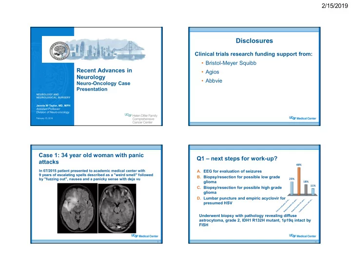
Case 1: 34 year old woman with panic attacks
3
In 07/2015 patient presented to academic medical center with 9 years of escalating spells described as a "weird smell" followed by "fuzzing out", nausea and a panicky sense with deja vu
Q1 – next steps for work-up?
- A. EEG for evaluation of seizures
- B. Biopsy/resection for possible low grade
glioma
- C. Biopsy/resection for possible high grade
glioma
- D. Lumbar puncture and empiric acyclovir for
presumed HSV
4
Underwent biopsy with pathology revealing diffuse astrocytoma, grade 2, IDH1 R132H mutant, 1p19q intact by FISH
E E G f
- r
e v a l u a t i
- n
- f
s e i . . . B i
- p
s y / r e s e c t i
- n
f
- r
p
- s
s . . . B i
- p
s y / r e s e c t i
- n
f
- r
p
- s
s . . . L u m b a r p u n c t u r e a n d e m . . .
23% 11% 18% 48%