Disclosures Nothing to disclose ABSTRACT PROFFERED TALK: Large ( 2 - - PowerPoint PPT Presentation

disclosures
SMART_READER_LITE
LIVE PREVIEW

Disclosures Nothing to disclose ABSTRACT PROFFERED TALK: Large ( 2 - - PowerPoint PPT Presentation

6/9/2017 Disclosures Nothing to disclose ABSTRACT PROFFERED TALK: Large ( 2 cm) Breast Cancers in Women Attending Regular Screening: Risk Factors and Implications for Prognosis FREDRIK STRAND, MD, MSc, PhD Cand. Karolinska Stockholm,


slide-1
SLIDE 1

6/9/2017 1

ABSTRACT PROFFERED TALK:

Large (≥ 2 cm) Breast Cancers in Women Attending Regular Screening: Risk Factors and Implications for Prognosis

FREDRIK STRAND, MD, MSc, PhD Cand.

Karolinska Stockholm, Sweden

Disclosures

  • Nothing to disclose

Personal introduction

  • Radiologist at Karolinska University

Hospital, Breast Imaging Unit

  • MSc in Engineering Physics
  • Epidemiological research on breast

cancer detection / non-detection (as PhD candidate)

  • Clinical research leading project on

deep learning techniques to improve breast cancer screening

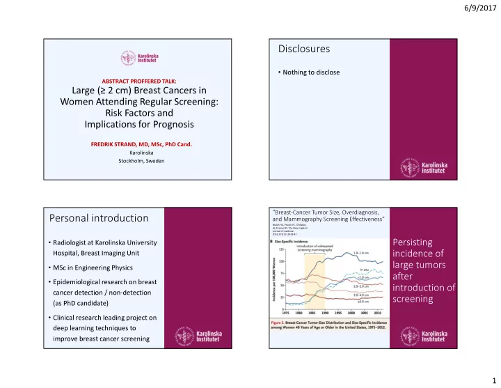
“Breast-Cancer Tumor Size, Overdiagnosis, and Mammography Screening Effectiveness”

Welch HG, Prorok PC, O'Malley AJ, Kramer BS. The New England journal of medicine. 2016;375(15):1438-47.

Persisting incidence of large tumors after introduction of screening

slide-2
SLIDE 2

6/9/2017 2

Source: Strand, unpublished

Without Screening

  • Larger Tumors

Larger tumor

  • Worse

prognosis Interval Cancer

  • Worse

prognosis

Screen-Detected Interval Cancers Non-Attenders

Source: Strand, unpublished

Large AND Interval

  • Worst

prognosis

slide-3
SLIDE 3

6/9/2017 3

The Study Large (≥ 2 cm) Breast Cancers in Women Attending Regular Screening: Risk Factors and Implications for Prognosis Authors

Fredrik Strand Johanna Holm Mikael Eriksson Sven Törnberg Edward Azavedo Per Hall Kamila Czene

Methods

Analysis Predisposing risk factors

  • Comparison: women with ≥ 2 cm vs. <

2 cm tumor at diagnosis

  • Overall and Stratified by detection

mode

Disease progression

  • Comparison: Age, BMI and PD
  • Stratified by detection mode

Study population

  • 2,358 cases of invasive breast cancer,

2001 to 2008 in Stockholm-Gotland

Predisposing factors known before diagnosis Purpose Improve screening

All Screen-detected Interval Cancers n=2,358 n=1,647 n=711 Large cancers 24% 20% 34% Interval cancers 30% 0% 100%

Results – predisposing risk factors for large (vs. small) tumor

Source: Strand, manuscript

slide-4
SLIDE 4

6/9/2017 4

Two different detection techniques – what hinders detection might not be the same

PALPATION (mainly) MAMMOGRAPHY MAMMOGRAPHY

Interval Cancer Screen-detected Cancer

Stratified by detection mode

Source: Strand, manuscript

“Post-dinner analysis” – potential confounders Education-level OR change from 2.46 to 2.47 Breast area (size proxy) OR change from 2.46 to 1.99

Source: Strand, manuscript

For Interval Cancers – tumor size at detection increases sharply with BMI

Probability of tumor remaining undetected until > 2 cm

Modell- estimated probabilities Source: Strand, manuscript

High BMI is related to aggressive tumor characteristics – especially for interval cancers

Molecular subtype by detection mode and BMI

slide-5
SLIDE 5

6/9/2017 5

Long-term follow-up

Does the association between BMI and adverse tumor characteristics translate into worse prognosis?

  • Disease Progression was defined as

either of local recurrence, distant metastasis or breast cancer death

  • We followed patients from date of

diagnosis (2001 to 2008) until the end of 2013

  • Possible survival bias since only

women who were alive at study entry in 2009 and 2010 could be included

(however – no bias identified when adjusting for year of study entry or time from diagnosis to study entry)

Among women with interval cancer - high BMI is associated with markedly worse prognosis

Disease progression = local recurrence, distant metastasis or breast cancer death

Conclusions

BMI as a predisposing risk factor

  • BMI increases probability of being

diagnosed with a larger tumor – especially among interval cancers Molecular subtype

  • Women with high BMI more often

have aggressive molecular subtypes

(especially Luminal B and Her2, not so much for basal)

Disease progression

  • Women with high BMI more often

experience disease progression (HR 2.0)

Women with high BMI

  • Women with high risk of mammographic masking

Supplemental ultrasound or MRI

PALPATION (mainly) MAMMOGRAPHY MAMMOGRAPHY

What to offer women with high BMI?

More frequent screening Also, take breast cancer risk into account

Less efficient for women with high BMI

Clinical Significance