Disclosures Update 2018 April 1-6, 2018 No relevant financial - - PDF document

disclosures
SMART_READER_LITE
LIVE PREVIEW

Disclosures Update 2018 April 1-6, 2018 No relevant financial - - PDF document

4/5/18 UCSF CME PRIMARY CARE MEDICINE: Disclosures Update 2018 April 1-6, 2018 No relevant financial relationship exists Common Upper Extremity Conditions You Will See in Office Practice Cindy J. Chang M.D. UCSF Primary Care Sports


slide-1
SLIDE 1

4/5/18 1

UCSF CME PRIMARY CARE MEDICINE: Update 2018

April 1-6, 2018

Common Upper Extremity Conditions You Will See in Office Practice

Cindy J. Chang M.D.

UCSF Primary Care Sports Medicine Associate Clinical Professor of Orthopaedics and Family and Community Medicine

Disclosures

No relevant financial relationship exists

Objective

Develop strategies to diagnose and manage common office problems including upper extremity injuries

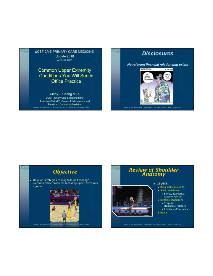
Review of Shoulder Anatomy

■ Layers ◆ Bony articulations (4) ◆ Static stabilizers ✦ Bones, ligaments,

capsule, labrum

◆ Dynamic stabilizers

✦ Scapular

stabilizers/rotators

✦ Rotator cuff muscles

◆ Bursa

slide-2
SLIDE 2

4/5/18 2

Shoulder Anatomy Shoulder Anatomy

Bony articulations

Shoulder Anatomy

Static Stabilizers

Shoulder Anatomy

Dynamic stabilizers

slide-3
SLIDE 3

4/5/18 3

Shoulder Anatomy

Dynamic stabilizers

Shoulder Anatomy

Dynamic stabilizers

■ Rotator Cuff

◆ Supraspinatus ◆ Infraspinatus uTeres minor uSubscapularis

Shoulder Anatomy

Dynamic stabilizers

■ Rotator Cuff

◆ Subscapularis ◆ (biceps tendon) ◆ Supraspinatus ◆ Infraspinatus uTeres minor

Shoulder Anatomy

Bursae

slide-4
SLIDE 4

4/5/18 4

Case #1

25 yo bike messenger was doored and fell off her bike, landing directly on the lateral aspect of her left shoulder

She was able to get back on her bike but 2 hours later, was in too much pain to continue working

3 days later after ice and NSAIDs she still had pain, difficulty lifting her arm

  • verhead or riding a bike

Based on this history, what is the least likely diagnosis?

  • 1. Clavicle fracture
  • 2. Rotator cuff tear
  • 3. AC joint sprain
  • 4. Subacromial bursitis/impingement

from traumatic contusion

  • 5. None of the above

Based on this history, what is the least likely diagnosis?

  • 1. Clavicle fracture
  • 2. Rotator cuff tear
  • 3. AC joint sprain
  • 4. Subacromial bursitis/impingement

from traumatic contusion

  • 5. None of the above

Clavicle Fractures

5% 5% 80% 80% 15% 15%

slide-5
SLIDE 5

4/5/18 5

Clavicle Fractures

◆ For any trauma with resultant loss of range of motion

and/or weakness and/or significant pain, get Xrays

Humerus Head Fractures

For any trauma with resultant loss of range of motion and/or weakness and/or significant pain, get Xrays ER view

AC Joint Sprain

◆ Deformity, crepitus, swelling, bruising of AC joint ◆ Pain with horizontal aDDuction ◆ Limited ROM and pain with resisted FF

, aBDduction

Shoulder Range of Motion

slide-6
SLIDE 6

4/5/18 6

Shoulder Range of Motion Shoulder Range of Motion

Shoulder Impingement Syndrome

Mechanism

■ Impingement under

acromion with flexion and internal rotation of the shoulder

■ Rotator cuff,

subacromial bursa and biceps tendon Symptoms

■ Pain with

◆ Overhead activities ◆ Sleep (Internal

rotation)

◆ Putting on a jacket

Shoulder Impingement Syndrome

Impingement of:

– Subacromial bursa – Rotator cuff muscles and

tendons

– Biceps tendon

Between

– Acromion – Coracoacromial ligament – AC joint – Coracoid process – Humeral head

Rotator cuff tendinosis

slide-7
SLIDE 7

4/5/18 7

Impingement Signs

Neer’s Sign

◆ Passive test –Arm fully pronated

and placed in forced flexion

–Trying to impinge

subacromial structures with humeral head

–Pain = Positive test

Sens = 83 % Spec = 51 % PPV = 40 % NPV = 89 %

MacDonald et al. J Shoulder Elbow Surg, 2000

Impingement Signs

Hawkins Sign

– Arm passively

forward elevated to 90 degrees, elbow is flexed, then shoulder forcibly internally rotated

– Trying to impinge

subacromial structures with humeral head

– Pain = positive test

Sens = 88 % Spec = 43 % PPV = 38 % NPV = 90 %

MacDonald et al. J Shoulder Elbow Surg, 2000

Always check cervical Impingement

■ Spurlings test for

cervical radiculopathy Sens = 64% Spec = 95% PPV = 58% NPV = 96%

Rotator Cuff Disease

■ Tendinitis ■ Partial thickness tear ■ Full (Complete)

thickness tear

■ May be due to:

◆ Impingement ◆ Degeneration ◆ Overuse ◆ Trauma

< 1% of shoulder injuries in persons < 30 yo are complete rotator cuff tears

slide-8
SLIDE 8

4/5/18 8 Rotator Cuff Disease

Etiology

◆ Overhead overuse

activities

◆ Trauma ◆ Instabililty ◆ Degenerative joint

disease

Pain radiates antlat and superior

◆ Often to deltoid insertion ◆ Often with night pain

Associated symptoms

◆ Giving way feeling ◆ Clicking, catching,

grinding

◆ Weakness

Rotator Cuff Disease

■ Painful Arc

◆ Pain with abduction

starting around 70 to 120

✦ Maximal at 90

◆ Pain with forward

flexion at 90-120

Can also be positive in impingement

Rotator Cuff Tendonitis/Tears

■ Subscapularis

◆ Lift-off test, Belly

press

■ Supraspinatus

◆ Empty can (Jobe test) ◆ Drop Arm Test

■ Infraspinatus, Teres

Minor

◆ Dropping sign ◆ Hornblower’s sign

Subscapularis Tests

■ Lift-Off Test

◆ Lift arm off the back ◆ If unable to maintain position

✦ Positive lift-off sign

◆ Make sure pt is not extending elbow ◆ Can then also test strength

Naredo et al. Ann Rheum Dis, 2002

For tendonitis: Sens = 50 % Spec = 84 % For tears: Sens = 50 % Spec = 95 %

slide-9
SLIDE 9

4/5/18 9

Subscapularis Tests

■ Belly-Press Test

◆ If unable to internally rotate arm to their back:

✦ Palms on belly ✦ Bring both elbows forward ✦ Resisted elbow forward flexion ✦ Not good to isolate superior fibers

Supraspinatus Tests

■ Jobe Test

◆ 30 deg anterior to

coronal plane

◆ Abduction 90 deg ◆ Thumbs up ◆ Isolate

supraspinatus muscle activity

◆ Resisted abduction

Supraspinatus Tests

Drop Arm Test Abducted arm slowly lowered

– May be able to lower

arm slowly to 90 (deltoid function)

– Arm will drop to side

if rotator cuff tear

Positive test

– patient unable to

lower arm further with control

– If able to hold at 90º,

pressure on wrist will cause arm to fall

Infraspinatus/Teres Minor Tests

Arms at the side

Elbows flexed

Resisted external rotation

For tendonitis: Sens = 57 % Spec = 71 % For tears: Sens = 36 % Spec = 95 %

Naredo et al. Ann Rheum Dis, 2002

slide-10
SLIDE 10

4/5/18 10

Infraspinatus/Teres Minor Tests

■ Dropping sign

◆ Infraspinatus ◆ Forearm is placed in 45

  • f external rotation

◆ Resist examiner’s hand ◆ If falls back to 0 of ER,

then + test

■ Hornblower’s sign

Teres minor and IS

If unable to stay ER when placed in 90/90 ER position, + test

Elbow rises above hand level when hand raised to mouth

Case #2

55 yo female with onset of right shoulder pain one year ago when playing tennis

◆ she is RHD

Had been “getting along” with it and controlling symptoms but began to notice gradual loss of motion despite ice and NSAIDs

Now presenting with pain all the time, including night pain, with inability to sleep on shoulder due to pain

She has had to buy new bras that clasp in front

What is your next step with this patient?

  • 1. Refer to PT if her ROM doesn’t improve with an

aggressive HEP at 1 mo F/U

  • 2. Control other comorbid conditions like HTN and

hyperlipidemia that predispose her to this problem

  • 1. Refer her to ortho for surgical manipulation under

anesthesia

  • 2. None of the above

What is your next step with this patient?

  • 1. Refer to PT if her ROM doesn’t improve with an

aggressive HEP at 1 mo F/U

  • 2. Control other comorbid conditions like HTN and

hyperlipidemia that predispose her to this problem

  • 3. Refer her to ortho for surgical manipulation under

anesthesia

  • 4. None of the above
slide-11
SLIDE 11

4/5/18 11

Adhesive Capsulitis

Spontaneous, gradual

  • nset of shoulder

stiffness and pain caused by tightening of joint capsule

70% female, 40-60 yoa

Comorbid conditions include diabetes, hypothyroid dz, RA

Usually occurs after shoulder immobilized or subconscious restricted motion after minor injury

Adhesive Capsulitis

■ IR/ADDuction first

to go and last to come back

■ Scapular

substitution

■ End range pain ■ Disuse atrophy

Natural History

■ 0-3 months gradual onset - painful ■ 2-9 months freezing ■ 4-12 months frozen ■ 5-26 months thawing ■ Usually self-limited Hannafin & Chiaia, Clin Orthop Rel Res, 2000

“The The art of

  • f medicine

co consists ts of amusing th the pa patient whi hile natur ure cu cure res th the disease.”

  • Vo

Voltaire

Treatment

■ Pain management (+/- sling) ■ Education and reassurance ■ Active home stretching

program

■ Physical Therapy ■ Oral NSAIDs (or steroids) ■ Glenohumeral injection-

capsular distension

■ Rarely needs surgery

(examination/manipulation under anesthesia or arthroscopic lysis of adhesions)

slide-12
SLIDE 12

4/5/18 12

Steroid injection?

■ RCT showed intraarticular steroid injection provided

better pain relief in the first 8 weeks than NSAIDs.

■ However, no difference seen in range of motion or

pain after 12 weeks

■ Results similar to other non-controlled studies Ranalletta M at al., Am J Sports Med, 2016

Case #3

40 yo male on vacation in Hawaii and crashed while surfing, and felt left shoulder pop out of joint when

  • underwater. Felt it pop back on its own seconds

later

Went to Urgent Care and got xrays and a sling. Controlled pain with ice and NSAIDS during the flight home.

Since then it clicks occasionally, and one week later is still cautious about moving his shoulder.

What would be accurate information about his injury?

  • 1. He likely had a posterior dislocation of his

shoulder

  • 2. He likely will have a positive relocation test
  • 1. If he is left-handed, he should get it

surgically stabilized

  • 2. A Beighton scale of 6 would be an indication

for surgery

slide-13
SLIDE 13

4/5/18 13

Shoulder Dislocation

Mechanism Anterior (>95%)

Force applied with shoulder in external rotation/ abduction Posterior (<5%)

Posterior force with shoulder in internal rotation/ adduction

EtOH (alcohol), Electrocution, Epilepsy

Diagnosis

Physical Exam

■ Tender anterior shoulder ■ May have decreased

sensation to deltoid

◆ axillary nerve

■ + Apprehension test ■ + Relocation test ■ + Sulcus sign (MDI)

XRay

Hill Sachs Lesion – compression fracture of posterior humerus Bony Bankart Lesion – Avulsion fracture of glenoid

MRI

Hill Sachs Lesion – compression fracture of posterior humerus Bankart Lesion – Avulsion of capsular attachment to the glenoid

slide-14
SLIDE 14

4/5/18 14

Recurrence vs. Age

%

Rowe CR. Prognosis in dislocation of the shoulder. J Bone Joint Surg Am, 1956

Complications after Dislocation

Acute rotator cuff tear

■ 40 to 60% incidence in patients > 40 years old

Frozen shoulder

■ Older the patient the stiffer they get

àmobilize early within 2-3 weeks

Recurrent dislocation

■ >90% recurrence < 20 years; 14% > 40 yrs

Treatment for Shoulder Instability

■ T – Traumatic ■ U – Unilateral ■ B – Bankart lesion ■ S – Surgical treatment

(refer for consultation)

■ A – Atraumatic ■ M – Multidirectional ■ B – Bilateral ■ R – Rehabilitation ■ I – Inferior capsular

shift

If Atraumatic Shoulder Instability…

Beighton hypermobility scale 5/9

slide-15
SLIDE 15

4/5/18 15

Little League Shoulder

Little League Shoulder – Imaging

  • Plain films show separation of proximal humeral

physis; get contralateral films if needed

  • Stop all throwing x 6 wks. Then gradual return to

throwing program. MRI if failing conservative tx x 3 months

Management of Shoulder Pain

Trauma

◆ Diagnosed/suspect capsule, labral, tendon, bony injury ◆ Ice/ACE wrap; Sling and NSAIDs for short duration ◆ Refer

✦ Surgery possible due to instab, mech symptoms,

complete RC tear, fracture

✦ If you are not comfortable with injections, fracture mgmt ✦ Not sure if imaging needed/helpful ✦ Patient request ■

Acute/Overuse

◆ Tendinitis, impingement, Grade 1-2 AC jt sprain ◆ Ice/ACE wrap; Sling and NSAIDs for short duration ◆ ROM, Stretches, foam roller, strengthening, posture,

x-train, internet

◆ Make a PT referral and f/u with you in 4-8 wks or after

4-6 PT visits

Thank you for your attention

slide-16
SLIDE 16

4/5/18 16

Nonspecific-type

Thoracic Outlet Syndrome

■ Refers to the largest group of TOS patients with

unexplained pain in the arm or both arms, scapular region, and cervical region

◆ Symptoms can begin after a traumatic event (eg, MVA) ◆ Growing population of students and workers who use

computers and smartphones all day

TOS Signs and Tests

■ Nonspecific TOS:

◆ Diffuse UE pain w/ or w/o guarding ◆ Nonfocal and non radicular findings ◆ Poor posture ◆ Tenderness over coracoid, pectoralis mm, scalenes;

tightness of mm

◆ Fullness in supraclav space from elevated rib ◆ Weakness and decreased sensation, tingling,

heaviness, fatigue, achiness

■ Adson’s maneuver ◆ Neck extended and rotated to

Affected side w/ Arm at side

while deeply inspiring and holding the breath, pulse checked

TOS Signs and Tests

■ Wright’s test – (Airplane)

◆ Affected arm abducted and

externally rotated, pulse checked, while taking a deep breath

■ Roos stress test – (Raise

the Roof)

◆ Shoulder abducted above the

head, externally rotated and repetitive opening and closing both hands into fists for at least 1 minute

■ Tests considered+ if

◆ reproduce symptoms ◆ decrease in pulse detected or

paresthesias for Adson or Wright