SLIDE 12 ◆12/18/16 ◆12
◆SCAD of the LAD Artery With Intramural Hematoma A 40-year-old woman presented 10 days
after a third-trimester miscarriage with troponin-positive non–ST-segment elevation myocardial infarction.
◆Katherine C. Michelis et al. JACC 2014;64:1033-1046
Longer-Term Management
■ Aspirin and beta blockade are the only nearly-
universal medications. Many get clopidogrel.
■ ACE-I for patients with LV dysfunction ■ Statins controversial; universal only for
patients with other indications
■ Cardiac rehabilitation geared towards SCAD
◆ Low weight threshold of 20 lbs ◆ Lower target heart rate and BP thresholds
■ Advise against continued hormone therapy in
those patients
■ Advise against future pregnancy (recurrence
rate thought to be high, but this is based on a 7-patient series)
Prognosis
■ In-hospital mortality relatively low, at under
5% in more-recent series
■ Recurrence rates are significant, with
recurrence in ~15% at 2 years and as high as 27% at 4 years (most recurrence is early).
■ In the largest prospectively-followed cohort of
280 patients, at a median followup of 2.3 years
◆ MACE was 20.4% ◆ Recurrent SCAD was 12.2%
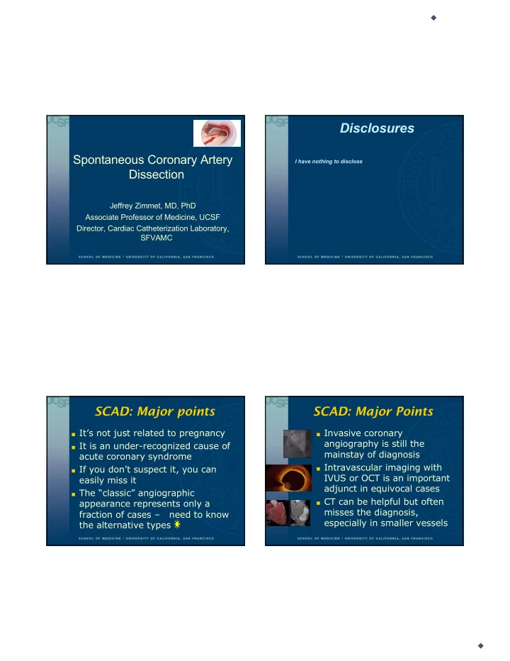
Wrap up
■
SCAD is an infrequent but increasingly-recognized cause of ACS
■
Preponderance in women, especially of younger age
■
Can occur in the peripartum period, although the majority of cases occur outside of this group
■
Also associated with physical and emotional stress, FMD, and with connective tissue disorders
■
Diagnosed by cardiac cath; often missed
■
Conservative therapy is favored when possible
■
Aspirin and beta blockade for short- and long-term therapy