disclosure surveillance for pulmonary vein stenosis Lars - - PDF document

disclosure
SMART_READER_LITE
LIVE PREVIEW

disclosure surveillance for pulmonary vein stenosis Lars - - PDF document

2017-03-10 disclosure surveillance for pulmonary vein stenosis Lars Grosse-Wortmann, MD, FRCPC gadolinium in young children Director, Cardiovascular MR The Hospital for Sick Children Toronto, Canada San Francisco, March 10, 2017 imaging


slide-1
SLIDE 1

2017-03-10 1

Lars Grosse-Wortmann, MD, FRCPC

Director, Cardiovascular MR The Hospital for Sick Children Toronto, Canada San Francisco, March 10, 2017

surveillance for pulmonary vein stenosis

gadolinium in young children

disclosure

  • pulmonary edema
  • discrete stenosis // diffuse hypoplasia
  • parenchymal changes, ground glass, mediastinal edema
  • flow redistribution
  • pulmonary hypertension (septal curvature, RVsp, mPAP, PA

flow profile)

  • attenuation of phasic flow
  • flow acceleration downstream

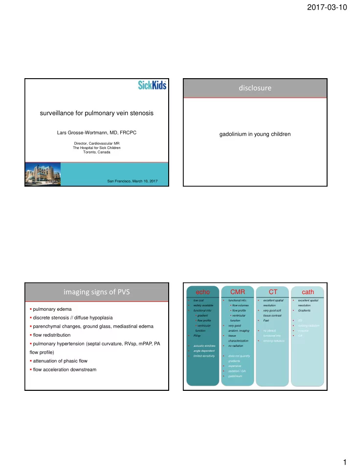
imaging signs of PVS

echo CMR CT

  • low cost
  • widely available
  • functional info:
  • gradient
  • flow profile
  • ventricular

function

  • RVsp
  • acoustic windows
  • angle dependent
  • limited sensitivity

cath

  • excellent spatial

resolution

  • very good soft

tissue contrast

  • Fast
  • no (direct)

functional info

  • ionizing radiation
  • functional info:
  • flow volumes
  • flow profile
  • ventricular

function

  • very good
  • anatom. imaging
  • tissue

characterization

  • no radiation
  • does not quantify

gradients

  • expensive
  • sedation / GA
  • gadolinium
  • excellent spatial

resolution

  • Gradients
  • 2D
  • ionizing radiation
  • invasive
  • GA
slide-2
SLIDE 2

2017-03-10 2

JCMR 2011

MRA

contrast-enhanced not ECG gated breathhold contrast-enhanced ECG gated respiratory navigated non-contrast ECG gated respiratory navigated

Grosse-Wortmann, JACC 2007 Grosse-Wortmann, JACC 2007

slide-3
SLIDE 3

2017-03-10 3 case 1: 4/12 ♂, AVSD, large PDA case 1: 4/12 ♂, AVSD, large PDA

PVRi (WU*m2) Qp/Qs FiO2 0.3 6.3 2.7 FiO2 0.6 6.1 3.1 FiO2 & NO 5.5

learning points

  • gradient may be overestimated with flow
  • full hemodynamic assessment, including Qp/Qs and

PVR, is helpful in decision making 1 month post-op 2 months post-op

4 months post-op stents in RUPV&LUPV dilation of RLPV&LLPV

1 month post-op 2 months post-op

4 months post-op stents in RUPV&LUPV dilation of RLPV&LLPV

learning points

  • PVS can progress rapidly
  • imaging of bioabsorbable stent feasible with CT
slide-4
SLIDE 4

2017-03-10 4 case 3: 3/12 ♀, s/p AVSD repair, LPV repair

learning points

  • PA (wedge) angiography ≠ direct PV injection
  • distinction between discrete narrowing and diffuse

hypoplasia can be challenging by cross-sectional imaging

  • recurrent chest infections
  • right pleural effusion
  • increased LLPV gradient

? RV/PA conduit exchange ? LLPV repair ? PAPVC repair ? APC occlusion?

► Qp/Qs, aortopulmonary collateral flow, PA flow distribution, RVEDV, RVEF

case 4: 2 years 10/12 ♀, RAI, AVSD, TGA, PA, mixed TAPVC, s/p AVSD repair, RV-PA conduit, sutureless repair RU+MPV learning points

  • decision making can be complex
  • CMR provides hemodynamic information that is

important for management

  • CMR can be performed without sedation / GA in young

children

  • total Qp/Qs = 1.7
  • no significant LLPV obstruction
  • RVEDVi 207ml/m2, RVEF 33%

case 4: 2 years 10/12 ♀, RAI, AVSD, TGA, PA, mixed TAPVC, s/p AVSD repair, RV-PA conduit, sutureless repair RU+MPV

+6 months

case 5: 9 months ♂, RUPV&RLPV stenosis

slide-5
SLIDE 5

2017-03-10 5

RPA LPA

learning points

  • PVS can be associated with mediastinal, perivascular

and peribronchial edema / lymphatic dilatation

  • PVS induces aortopulmonary collaterals
  • ipsilateral PA flow can be reduced and net flow can be

retrograde

case 5: 9 months ♂, RUPV&RLPV stenosis

case 6: 16 months ♂, prematurity, BPD, hemoptysis

? mediastinal hemangioma

learning points

  • prematurity and bronchopulmonary dysplasia are risk

factors for development of PVS

  • the obstructed PV can be decompressed via
  • PV-to-PV collaterals
  • PV-to-systemic venous collaterals

case 7: 16 months ♂, prematurity, BPD, hemoptysis

slide-6
SLIDE 6

2017-03-10 6

Grosse-Wortmann, Cardiol Young 2007

case 8: 5 year ♀, RAI, unbalanced AVSD, DORV, PS, TAPVC, s/p TAPVC repair & bilateral BCPC

learning points

  • PVS can be associated with development of massive

aorto-pulmonary collaterals, especially in patients with BCPC or Fontan circulations

MRI 1 MRI 2 MRI 3 RPA flow (l/min/m2) .93 1.18 3.47 LPA flow (l/min/m2) .73

  • .33
  • 3.06

LLPV flow (l/min/m2) 1.01 .1 .02 Qp/Qs .63 .66 1.76

Grosse-Wortmann, Cardiol Young 2007

case 9: 5 year ♀, RAI, unbalanced AVSD, DORV, PS, TAPVC, s/p TAPVC repair & bilateral BCPC

diagnosis

surveillance

echo, x-sectional echo, x-sectional echo, x-sectional

3-6 months 12 months

progression surgery / cath

1 month

consider palliation or HLTx

CT or MRI ?

  • default = MRI
  • CT
  • ?stent [cath]
  • comparison with previous CT
  • functional status known or not relevant for mx
  • GA for MRI to be avoided
  • renal failure
  • airway // pulmonary parenchymal // mediastinal

imaging

slide-7
SLIDE 7

2017-03-10 7 summary

  • clinical question  choice of imaging modality
  • echo does not reliably r/u PVS
  • MRI angiography: ECG gated, contrast-enhanced
  • CT angiography: preferably ECG gated
  • MRI can be performed without sedation / GA in

infants and preschool children

  • cath may be needed to determine patency

future directions

► screening by echocardiography for select patients ► image fusion ► non-contrast MR angiography ► predictive value of imaging biomarkers ► role of exercise echocardiography & CMR

Shi-Joon Yoo Rachel Vanderlaan Christopher Caldarone Andreea Dragulescu Andrea Wan Hartmut Grasemann Tilman Humpl Jennifer Russell

save the date