
SLIDE 1
Page
Diffusion Tensor Imaging of Mild Diffusion Tensor Imaging of Mild - - PowerPoint PPT Presentation
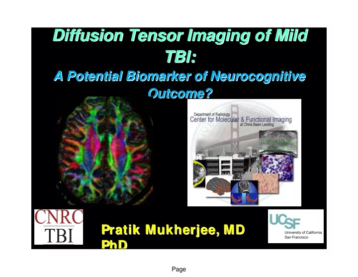
Diffusion Tensor Imaging of Mild Diffusion Tensor Imaging of Mild TBI: TBI: A Potential Biomarker of Neurocognitive Neurocognitive A Potential Biomarker of Outcome? Outcome? Pratik Mukherjee Mukherjee, MD , MD Pratik PhD PhD Page
Page
Page
Page
Page
Page
Page
Page
– All with GCS 13-15 in the Emergency Dept.; no prior history of head trauma – All with loss of consciousness; none for more than 30 min. – All with post-traumatic amnesia
» Positive for brain injury
» Positive for hemorrhagic axonal shearing injury
» Positive for non-hemorrhagic axonal shearing injury
» Positive for cerebral contusions
Lee H, Wintermark M, Gean A, Ghajar J, Manley G, Mukherjee P. J Neurotrauma 2008; 25:1049-56
Page
Page
Lee H, Wintermark M, Gean A, Ghajar J, Manley G, Mukherjee P. J Neurotrauma 2008; 25:1049-56
Page
Page
– Is sensitive to microstructural changes within white matter tracts, which may improve the detection of axonal injury improve the detection of axonal injury Arfanakis Arfanakis et al. et al. AJNR AJNR (2002) and many other studies (2002) and many other studies – can localize axonal shearing injury to specific white matter tracts, for structure structure-
function correlation Le et al. Le et al. Neurosurgery Neurosurgery (2005) and other studies (2005) and other studies – can provide quantitative quantitative pathophysiological pathophysiological information information that might be useful for determining prognosis and monitoring therapeutic interventions in TBI Huisman Huisman et al. et al. AJNR AJNR (2004) and other studies (2004) and other studies – – 3T MRI with parallel imaging 3T MRI with parallel imaging vastly improves the ability to perform high-resolution, high quality DTI in a clinically feasible scan time
Page
Pure water at 37˚C: ADC ~ 3.0 x 10⁻³ mm2/sec Normal adult brain: (GM & WM) ADC ~ 0.7 x 10⁻³ mm2/sec
in the range b = 0 - 1000 sec/mm2 …
Normal term newborn brain: GM: ADC ~ 1.1 x 10⁻³ mm2/sec WM: ADC ~ 1.5 x 10⁻³ mm2/sec Fractional Anisotropy (FA): 0 (spherical) to 1 (linear)
Page
min)
min)
(5 min)
1.8-mm TE=64 ms, TR=14 s, 55 diffusion-encoding directions, b=1000 s/mm2 ASSET parallel imaging with acceleration factor of 2 (13 min)
Page
centrum semiovale superior longitudinal fasciculus cingulum bundle corpus callosum, body
Page
pyramidal tract decussation, superior cerebellar peduncle cingulum decussation, middle cerebellar peduncle anterior commissure transverse pontine fibers middle cerebellar peduncle ILF SLF corpus callosum
Page
Page
Reaction Time versus # of microbleeds
R = -.08, p=0.701
400 500 600 700 800 900 1000 1100 5 10 15 20 # of traumatic microbleeds (conventional MRI) Mean RT (ms)
Page
Page
Reaction Time affected by Diffuse Axonal Injury
R = 0.49, p=0.012
400 500 600 700 800 900 1000 1100 1 2 3 4 5 6 7 8 Number of DTI lesions Mean RT (ms)
Page
Page
FACT – fiber assignment by continuous tracking in 3D along the primary eigenvector (Mori et al. 1999)
dense seeding – multiple seed points within a voxel (Conturo et al. 1999; Mori et al. 1999) multi-ROI filtering – retain only those tracts passing through start and end ROIs, and other intermediary ROIs (Conturo et
interpolation – step sizes smaller than a voxel (Conturo et al. 1999)
Page
Page
Page
Page
Bilateral UNC correlates with Memory in Normal Adults R = 0.52
4 6 8 10 12 14 16 18 0.4 0.45 0.5 0.55 0.6 0.65 0.7
FA of UNC LDFR Memory Score from CVLT
Left hemisphere ACR correlates with Conflict in Normal Adults R = -0.42
25 45 65 85 105 125 145 165 185 0.4 0.45 0.5 0.55 0.6
FA of ACR Conflict Score from ANT (ms)
Page
Bilateral Uncinate Fasciculus correlates with Memory in mild TBI R = 0.518
2 4 6 8 10 12 14 16 18 0.3 0.4 0.5 0.6 0.7 0.8
FA of UNC LDFR Memory Score from CVLT
Left hemisphere Anterior Corona Radiata correlates with Conflict in mild TBI R = -0.47
50 100 150 200 250 300 350 0.2 0.4 0.6 0.8 1
FA of ACR Conflict Score from ANT (ms)
Page
Page
Control Control vs vs TBI TBI (p) (p)
Left 0.492±0.0 22 0.474±0.21 0.475±0.02 0.472±0.0 21 0.00 9 0.01 5 0.00 6 Right 0.470±0.0 22 0.455±0.24 0.459±0.02 0.457±0.0 24 0.03 3 0.08 0.06 5 Average 0.481±0.0 20 0.465±0.02 0.467±0.01 8 0.464±0.0 18 0.00 8 0.01 9 0.00 9
Page
Control Control vs vs TBI TBI (p) (p)
Left 0.550±0.0 24 0.539±0.02 3 0.543±0.02 4 0.535±0.0 21 0.11 1 0.34 2 0.04 5 Right 0.533±0.0 22 0.524±0.02 3 0.521±0.02 3 0.518±0.0 23 0.15 8 0.06 3 0.03 6 Average 0.542±0.0 22 0.531±0.02 0.532±0.02 1 0.527±0.0 19 0.10 6 0.13 5 0.02 6
Page
Control Control vs vs TBI TBI (p) (p)
Left 0.562±0.0 33 0.549±0.02 8 0.553±0.03 6 0.542±0.0 31 0.18 6 0.36 8 0.05 9 Right 0.520±0.0 22 0.512±0.02 5 0.515±0.02 9 0.510±0.0 23 0.26 3 0.46 3 0.14 3 Average 0.541±0.0 26 0.531±0.02 5 0.534±0.03 1 0.526±0.0 23 0.18 0.37 9 0.06
Page
Control Control vs vs TBI TBI (p) (p)
Left 0.516±0.0 22 0.517±0.01 9 0.513±0.02 3 0.516±0.0 18 0.92 7 0.64 3 0.97 8 Right 0.491±0.0 22 0.484±0.02 7 0.480±0.02 6 0.482±0.0 25 0.63 5 0.13 1 0.23 3 Average 0.504±0.0 18 0.505±0.02 1 0.500±0.02 1 0.503±0.0 19 0.83 8 0.13 1 0.87 2
Page
Control Control vs vs TBI TBI (p) (p)
Left 0.587±0.0 28 0.589±0.02 2 0.586±0.02 1 0.581±0.0 24 0.76 2 0.94 5 0.47 9 Right 0.573±0.0 23 0.571±0.02 7 0.570±0.02 1 0.561±0.0 22 0.74 8 0.72 5 0.11 9 Average 0.580±0.0 24 0.580±0.02 2 0.578±0.02 3 0.571±0.0 22 1.00 0.82 9 0.23 9
Page
Control Control vs vs TBI TBI (p) (p)
0.556±0.0 22 0.542±0.02 2 0.544±0.02 5 0.542±0.0 19 0.03 8 0.08 4 0.03 2
Page
Control Control vs vs TBI TBI (p) (p)
0.669±0.0 25 0.658±0.02 7 0.656±0.03 0.653±0.0 27 0.14 4 0.09 4 0.04 4
Page
– Growing increasingly sensitive to the focal lesions of mild TBI, especially microbleeds of TAI on T2* GRE or SWI – However, no evidence that focal lesions are relevant to long-term neurocognitive status or functional recovery in mild TBI
– DTI measures such as FA are correlated with cognitive processing speed, memory, & attention – Specific (micro)structure-function relationships can be found between particular white matter tracts and their associated neurocognitive domain (UF-memory, ACR-attention) – Reduced microstructural integrity of specific WM tracts can be detected within 2 wks after mild TBI – prognostic biomarker? – However, overlap with normal variation may limit utility for clinical diagnosis in individual cases of mild TBI
Page
– DTI is sensitive in blunt trauma, so might also be sensitive in blast – Does the distribution of microstructural WM injury differ in blast?
» Blunt TBI: anterior WM tracts (prefrontal connectivity) appear most affected » Blast TBI: are posterior cerebral and posterior fossa tracts most affected?
– What is the relationship between white matter FA and biomechanical susceptibility to blast injury?
» Current limitation of DTI: it is largely unknown what are the biological determinants of DTI parameters such as FA, and what are the pathophysiological changes that cause reduced FA after trauma
– Can DTI be used to help model strain and deformation in the brain due to blast exposure?
Page
Cornell Cornell Sumit N. Niogi, PhD Bruce McCandliss, PhD UCSF @ SFGH UCSF @ SFGH Geoffrey T. Manley, MD PhD Alisa Gean, MD Hana Lee Michele Meeker, RN Supported by the a collaborative grant from the James S. McDonnell Foundation administered through the Brain Trauma Foundation (Cornell, UC Berkeley, UCSF) UCSF Radiology UCSF Radiology Joshua Ng Srivathsa Veeraraghavan Daniel B. Vigneron, PhD Michael Wahl Duan Xu, PhD Brain Trauma Foundation Brain Trauma Foundation Jam Ghajar, MD PhD