content
play

Content Visual Function Endpoints in Clinical Trials - PDF document

Content Visual Function Endpoints in Clinical Trials


  1. Content Visual Function Endpoints in Clinical Trials �������������� ���� ������ �������������������������������������� �������� Clinical View � Basics: Structure and Function � Function Testing: Psychophysics in Ophthalmology Eberhart Zrenner � Function Testing Electrophysiology in Ophthalmology � Retinal Imaging Center for Ophthalmology � Assessing Activities of Daily Living Institute for Ophthalmic Research University of Tübingen � Patient Reported Outcomes (PRO) of Visual Function Germany � Examples from Ongoing Studies Sclera � Financial Disclosure Choroid Cornea RPE � The author performs or performed advisory tasks during Iris Retina the 5 most recent years for Acucela, Allergan, Bayer, Lens Boehringer Ingelheim, Merck, Neurotech, Pfizer, Retina Fovea,Macula Implant AG, Servier and QLT; he is shareholder of Retina Implant AG and inventor on patents concerning electronic subretinal implants.
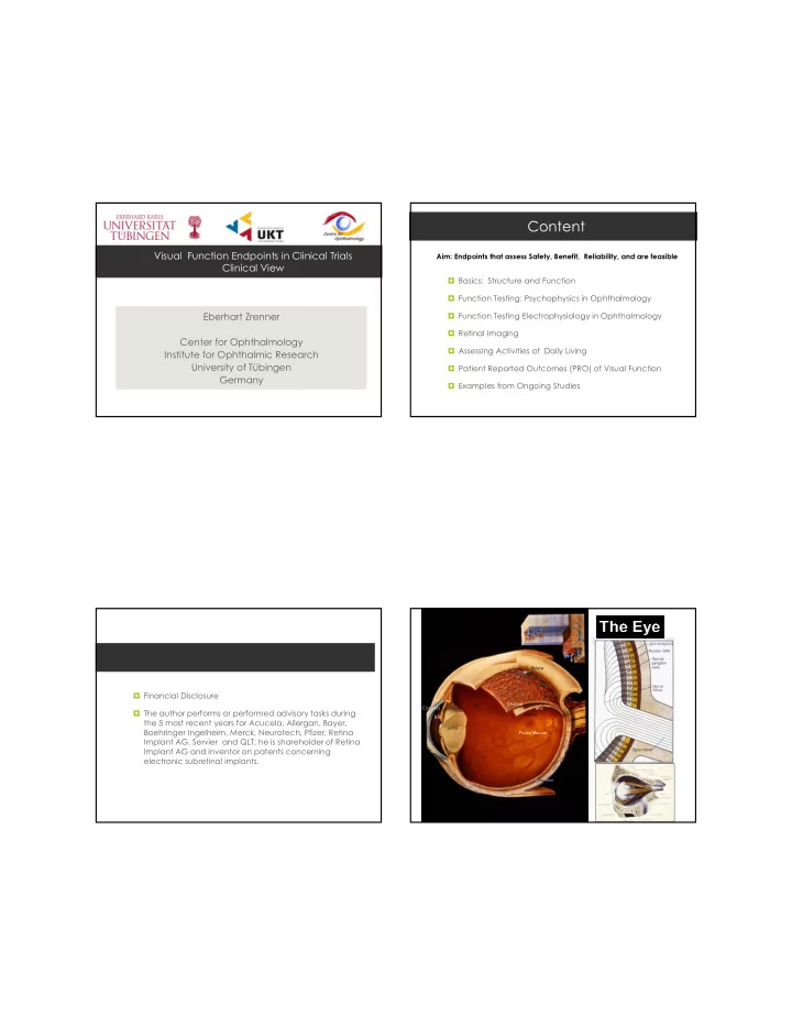
  2. Retinal Structure and Function The Phototransduction process: many things can go wrong ���� �������������� ������������������ Low sensitivity High sensitivity Rhodopsin High spatial resolution Low spatial resolution Colour discrimination No colour discrimination ��� �������� ����������� Information Processing in the Retina The Short, Middle and Long Wavelengths Sensitive Cones Cone Mosaic in the Fovea ~ 120 million photoreceptors ⇓ ⇓ ⇓ ⇓ „The natural „Darkness Channel“ computer“ Courtesy of T. Sharpe „Brighness Channel“ ⇓ ⇓ ⇓ ⇓ Courtesy of H. Waessle cone ~ 1 million ganglion cell fibres sending rods From: J. Dowling: The Retina. 1987 pulses to the brain ( courtesy of Paul R. Martin, 1998 )

  3. ������� ���������� ������������������������ Functional Diagnostics Best Corrected Visual Acuity (BCVA) Visual Acuity, Refraction, Anterior segment Reading speed, Pupil, Eye movement, � Snellen chart Examination intraocular pressure Fundi oculi ��� Visual fields ERG multifocal ERG Colour vision ��� ���� Cones ��� ��� ��� ��� ��� ���� Functional Diagnostics Functional Diagnostics Best Corrected Visual Acuity (BCVA) Best Corrected Visual Acuity (BCVA) � Snellen chart � Snellen chart � ETDRS chart (Standard) � ETDRS chart (Standard � Freiburg Visual acuity Test (FRACT) with Landolt CBring (ComputerBScreen test ) K. SchulzeBBonsel, et al., Visual Acuities “Hand Motion” and “Counting Fingers” Can Be Quantified with the Freiburg Visual Acuity Test. ��������������������������

  4. Functional diagnostics FunctionalDiagnostics Colour vision testing Contrast Vision � PelliBRobson Chart � ������ �������� ����� ����� ���� � Mesoptometer � Lanthony Panel DB � With and without glare 15 Test � Hamilton Veale Test � Roth 28Bhue Test � FarnsworthBMunsell 28 or 100 Test WEB based evaluation tool: http://www.torok.info/colorvision/dir_for_use.htm (Dr. Bela Török) achromat � ������������ protan N deutan Functional diagnostics Functional diagnostics Visual Field testing Visual Field testing � Visual field testing � Visual field testing � Static perimetry 30° � Static perimetry 30° � Kinetic perimetry 90° � Kinetic perimetry 90° � Microperimetry Central Vision Eye Movement Compensated

  5. Functional Diagnostics Functional diagnostics: Pupillography Dark Adaptation Adaptometer � Relative Afferent Pupillary Defect (RAPD) (Roland Consult) Infrared Image � Pupillography cone branch rod branch AMTECH Pupillograph ������� / Surrogate Endpoints ������������� � A ��������� �������� , according to the FDA is a biomarker that is “reasonably likely, based on epidemiologic, therapeutic, pathophysiologic, or other evidence to predict clinical benefit.” The best surrogate endpoint is a biomarker that changes along with clinical endpoints. ���!��������"�������������������������� ��������������������������������#����� ���$������������%������ "�#����� ��&������'"�'�������������"�'����������������������(����������)�����*+,-+���������������������*..+ Csaky et al, IOVS 2009

  6. Categorization of function tests: Clinical Electrophysiology of the Visual System Electroretinogram: s ites of action /( ��� ������������� ��� &��0( 1������� ���2�� ����������&1��( *( %���� ������ 3��������� ��0�&����0( ����������0�&���0( ��� ������������ ��� &�%0( -( $����������� "�� 34����� ��� ���� ��� �� ��� 5 6� ������ ������� �������� ������������������������ �� ������ �������� ��7���� ��� ������ ���!��������"�������������������������� ��������������������������������#����� ���$������������%������"�#����� �� &������'"�'�������������"�'����������������������(����������)�����*+,-+���������������������*..+ Outer Retina: ISCEV Standard Ganzfeld ERG Lublin Oct. 2005 Origin of the ERGBresponses DTLBFibre Electrode ERG b-wave 1. Isolated rod responses OP´s 2. Rod and cone Max. Responses ERG a-wave Jet Electrode Horizontal cells Ganglion cells Photoreceptors Bipolar cells 3. Inner Retina OPs 4. Isolated Cone Responses 8�7 ���������� 9 5. Cone flicker Responses �����3������������������������� �������� ������������� ����� Retinal pigment epithelium &*..+�������(��:���%��������� *..;<//+�=;>?? Choriod capillaries Amacrine cells Bruch's membrane Müller glia cells For further Information see www.iscev.org

Download Presentation
Download Policy: The content available on the website is offered to you 'AS IS' for your personal information and use only. It cannot be commercialized, licensed, or distributed on other websites without prior consent from the author. To download a presentation, simply click this link. If you encounter any difficulties during the download process, it's possible that the publisher has removed the file from their server.

Recommend


More recommend