
CASE REPORT
12 | TSMJ 2016
Concealed Accessory Pathway in Late Presentation Wolff-Parkinson-White Syndrome
Stephanie Rose1, Richard Armstrong2, David Moore2
1Third year medicine, Trinity College Dublin 2Department of Cardiology, Adelaide and Meath Hospital, Dublin, incorporating the National Children’s Hospital
Background
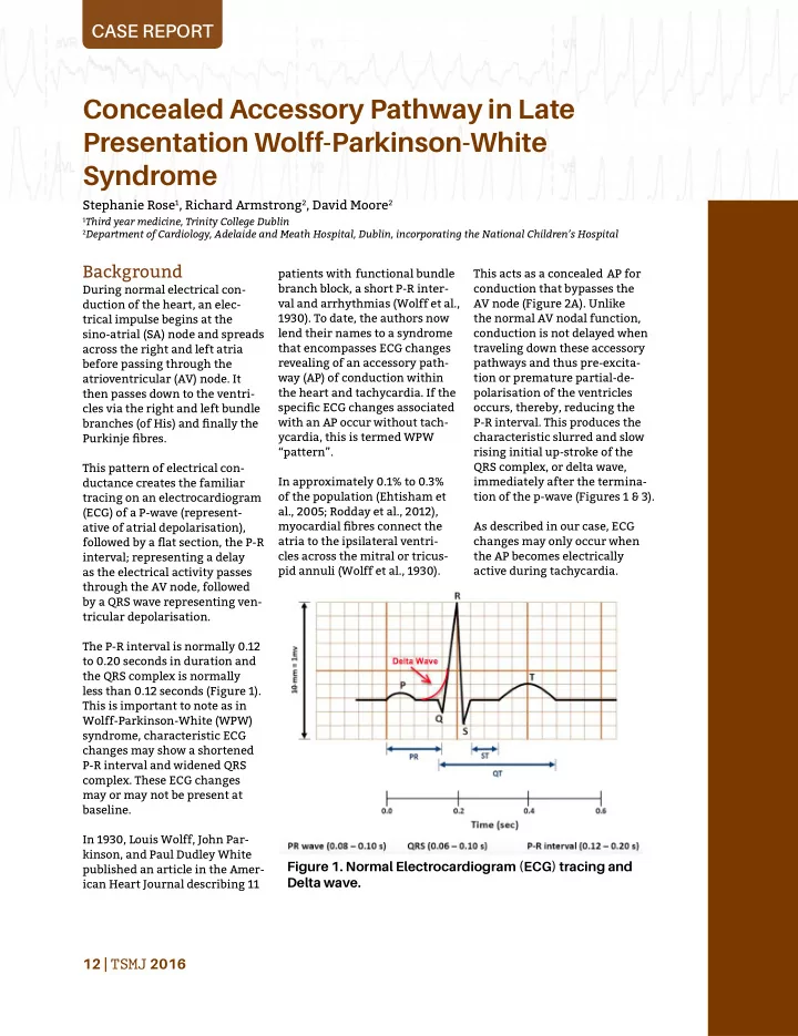
During normal electrical con- duction of the heart, an elec- trical impulse begins at the sino-atrial (SA) node and spreads across the right and left atria before passing through the atrioventricular (AV) node. It then passes down to the ventri- cles via the right and left bundle branches (of His) and finally the Purkinje fibres. This pattern of electrical con- ductance creates the familiar tracing on an electrocardiogram (ECG) of a P-wave (represent- ative of atrial depolarisation), followed by a flat section, the P-R interval; representing a delay as the electrical activity passes through the AV node, followed by a QRS wave representing ven- tricular depolarisation. The P-R interval is normally 0.12 to 0.20 seconds in duration and the QRS complex is normally less than 0.12 seconds (Figure 1). This is important to note as in Wolff-Parkinson-White (WPW) syndrome, characteristic ECG changes may show a shortened P-R interval and widened QRS
- complex. These ECG changes
may or may not be present at baseline. In 1930, Louis Wolff, John Par- kinson, and Paul Dudley White published an article in the Amer- ican Heart Journal describing 11 patients with functional bundle branch block, a short P-R inter- val and arrhythmias (Wolff et al., 1930). To date, the authors now lend their names to a syndrome that encompasses ECG changes revealing of an accessory path- way (AP) of conduction within the heart and tachycardia. If the specific ECG changes associated with an AP occur without tach- ycardia, this is termed WPW “pattern”. In approximately 0.1% to 0.3%
- f the population (Ehtisham et
al., 2005; Rodday et al., 2012), myocardial fibres connect the atria to the ipsilateral ventri- cles across the mitral or tricus- pid annuli (Wolff et al., 1930). This acts as a concealed AP for conduction that bypasses the AV node (Figure 2A). Unlike the normal AV nodal function, conduction is not delayed when traveling down these accessory pathways and thus pre-excita- tion or premature partial-de- polarisation of the ventricles
- ccurs, thereby, reducing the
P-R interval. This produces the characteristic slurred and slow rising initial up-stroke of the QRS complex, or delta wave, immediately after the termina- tion of the p-wave (Figures 1 & 3). As described in our case, ECG changes may only occur when the AP becomes electrically active during tachycardia.
Figure 1. Normal Electrocardiogram (ECG) tracing and Delta wave.