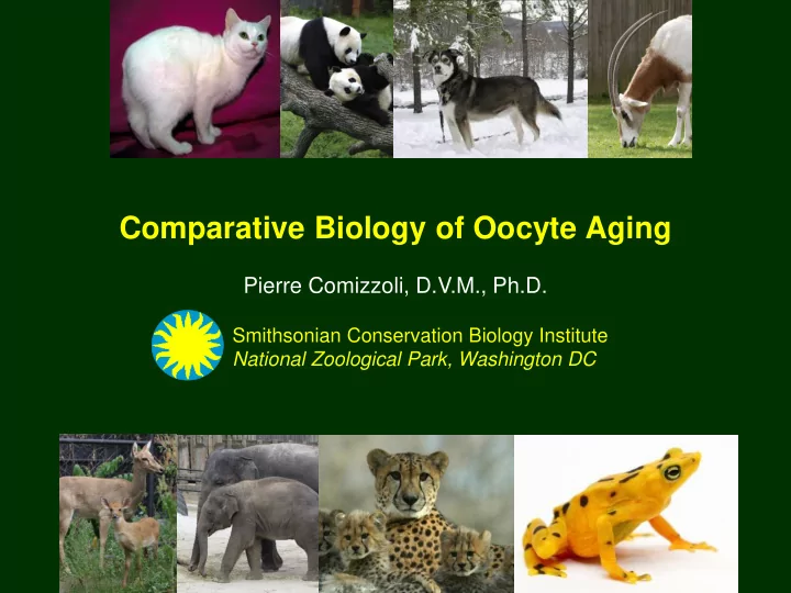

Comparative Biology of Oocyte Aging Pierre Comizzoli, D.V.M., Ph.D. Smithsonian Conservation Biology Institute National Zoological Park, Washington DC
Disclosure I have nothing to disclose
Contribution of Reproductive Science and Gamete Biology to Conservation Biology Value of basic/comparative studies Progestagens (ng/ml) 1.2 0.8 0.4 0.0 0 250 500 750 1000 Days → Scholarly knowledge and conservation actions in situ and ex situ (enhancing natural mating, maintaining genetic diversity) → Development of Assisted Reproductive Techniques and Genome Resource Banking (overcoming mating difficulties, preserving fertility, maintaining genetic diversity)
Diversity and Complexity of Reproduction and Gamete Biology Only few species well described among 5,400 mammal species As many mechanistic differences in reproduction as there are species Diversity in fertility issues (aging, teratospermia , sensitivity to stress…) Species-specificities in hormonal stimulations and in vitro culture conditions Difficult to compare human and animal reproductive aging (different lifespan) No ‘menopause’ in animals but age-related loss of fecundity
Diverse Reproductive Physiologies Seasonal Estrous Cycle Estrus (days) Ovulation Gestation Breeder (days) (days) Black-footed 5 – 29 2 – 9 63 – 71 No Ind. cat 5 – 10 50 – 70 Bobcat Yes 44 Ind. 7 – 23 2 – 6 90 – 98 Cheetah No Ind. Clouded 25 – 30 3 – 6 85 – 93 Yes Spont. leopard 14 – 21 3 – 7 64 – 67 Domestic cat Varies Both Asian 100 – 110 2 – 4 640 – 660 No Spont. elephant Eld’s deer 20 – 24 1 – 2 230 – 240 Yes Spont. 7 – 10 80 – 180 Giant panda Yes 1 Spont.
Values of Comparative Folliculogenesis and Oocyte Biology Animal models are essential to improve our understanding of aging mechanisms and develop mitigation strategies Limitations of existing laboratory models because of size, anatomy, and physiology (including lifespan) Differences in ovarian anatomy and histology
Values of Comparative Folliculogenesis and Oocyte Biology Differences in oocyte size (minimal), nucleo-cytoplasmic ratio, lipid content Rodents Bovids Suidae Felids Human Oocyte diam. 80 110 125 110 110 ( µm) Germ. vesicle 30 35 35 40 40 diam. ( µm ) In vitro <24 hr ~24 hr ~44 hr ~28 hr ~24 hr maturation Differences in folliculogenesis (timing, follicle waves, polyovulations) Differences in oocyte competence related to the follicular size Differences in GV competence related to the follicular size
Reproductive Aging: Complex and Multifactorial Mechanisms Mainly described in human Aging of organism affecting folliculogenesis and ovulation (hormone level changes) Egg quantity and quality significantly declines with reproductive age (>35 yr) Increase in miscarriages, infertility, and birth defects Changes in reproductive tract affecting conception, embryo development, implantation, and pregnancy Accumulations/exposures during reproductive life: Irreparable damage, long arrest at the GV stage, increased oxidative stress during folliculogenesis
Oocyte Aging: Complex and Multifactorial Mechanisms Too! Changes in: GV chromatin configuration and integrity GV epigenetics/transcriptomics GV proteomics Cytoplasm (mitochondria number and function, protein metabolism) Zona pellucida Connections with cumulus cells Ovarian environment (fibrosis) As a result: Defects in meiotic maturation (chromosome segregation, aneuploidy) Defects in fertilization, embryo development, implantation, pregnancy Need systematic approaches with proper models for each aspect
Chromatin Configuration and Integrity Chromatin configuration and competence in the cat model A B C D E F G H ~8% of oocytes with abnormal configuration and DNA damage at any age ~10% in adult ungulates vs. 25% in old individuals (past 14 yr)
Germinal Vesicle Epigenetics Aging mouse oocyte - Decrease in expression of histone deacetylases (HDAC) and DNA methyl-transferases (DNMT) Genome-wide DNA methylation is lower Histones are more acetylated Key histone methylations are altered Need for alternate models (closer to human in size and timing)
Germinal Vesicle Epigenetics Distribution of Histone Deacetylase 2 during folliculogenesis and transcriptional silencing in the cat model Translocation occurs earlier in older individuals (>12 year)
Manipulation of Epigenetics in Germinal Vesicles Reversible and global de-acetylation to mitigate aging Control 0.5 mM resveratrol 1.0 mM resveratrol 1.5 mM resveratrol
Germinal Vesicle Epigenetics Primary regulations of histone methylations during folliculogenesis in the cat model Primary regulations of histone methylations are modified in old cats (>12 year)
Changes in Ovarian Environment Squirrel monkey Cattle Cheetah
Aging Study in Cheetahs ( Acinonyx jubatus ) A Counterexample Drop of fertility in older females No pregnancies after natural breeding in old females Normal ovarian cycles Good ovarian response to exogenous gonadotropins But no conception after intra-uterine artificial insemination
Aging Study in Cheetahs ( Acinonyx jubatus ) 1.8 200 Estradiol 180 1.6 Progestagens 160 Oocyte 1.4 aspiration Progestagens (µg/g dry feces) 140 Estradiol (µg/g dry feces) 1.2 120 1.0 hCG 100 0.8 eCG 80 * * 0.6 * * 60 0.4 40 0.2 20 0.0 0 Day
Aging Study in Cheetahs ( Acinonyx jubatus ) Comparison of ovarian anatomy Oocyte aspiration and IVF Percentages of fertilization and embryo development were not different between young and old females Oocyte quality is not affected by the age of the female (including microtubules, mitochondrial functions …)
Aging Study in Cheetahs ( Acinonyx jubatus ) Increase of uterine wall thickness in old females High prevalence of cystic endometrial hyperplasia
Age Effect in Eld’s Deer ( Rucervus eldii thamin ) In vitro maturation In vitro fertilization and culture for 7 days in deer SOF medium
Age Effect in Eld’s Deer ( Rucervus eldii thamin ) Adapted hormone treatments to stimulate folliculogenesis in old donors Less embryo development and no pregnancy with donors >10 yr old Heat stress > aging - Older females are more sensitive
Lessons Learned Have induced ovulator less oocyte aging issues? (higher production rate of oocytes) Ungulates seems to be more prone to effect of age Understanding oocyte aging through methods used for mitigations (optimization and development of new tools): Adapted hormone stimulation in aged patients (deer) Resveratrol exposure in cat oocytes Germinal vesicle transfer (cytoplasmic aging > nuclear aging)
Take-Home Messages Values of comparative studies to advance knowledge on oocyte aging and ‘shed a new light’ on human fertility studies Difficult to compare oocyte aging in animal species (no menopause, uterine pathology or heat stress are prevalent) We know very little about oocyte aging in animal species Elephants could be an excellent model but no knowledge on the oocyte Systems biology will help to better understand and mitigate oocyte aging
Recommend
More recommend