
SLIDE 1
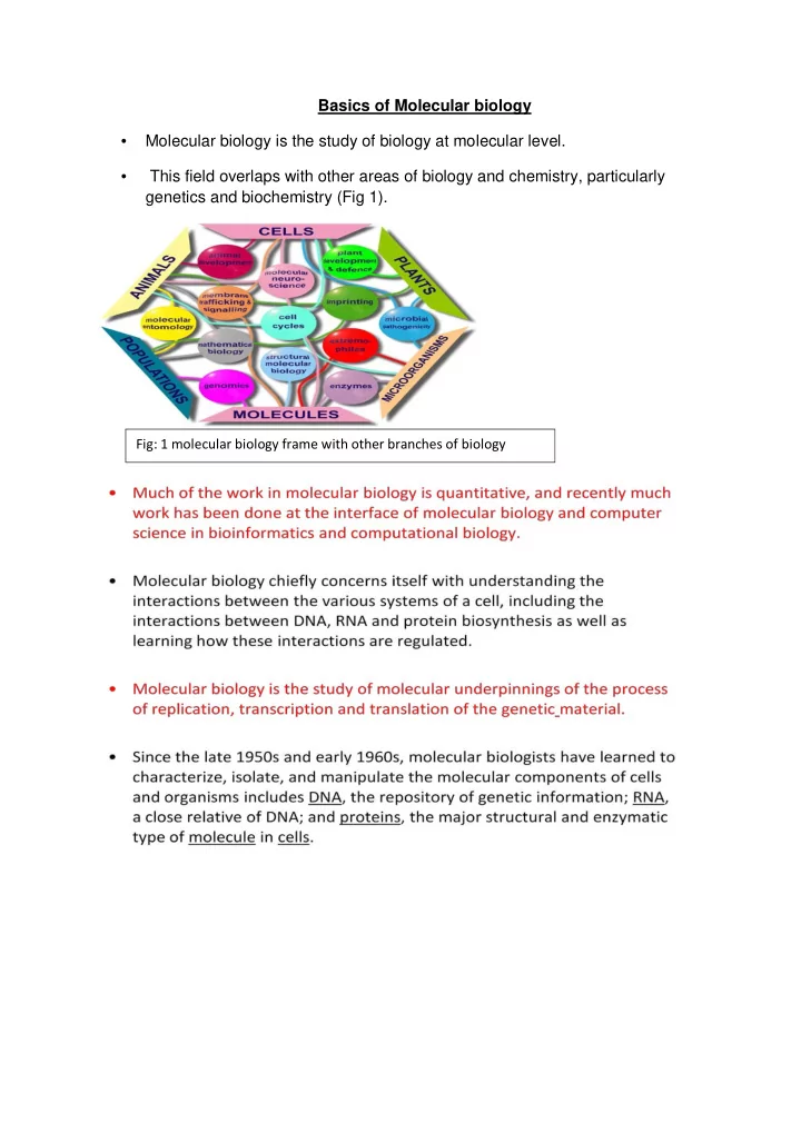
- Molecular biology is the study of biology at molecular level.
Basics of Molecular biology
- This field overlaps with other areas of biology and chemistry, particularly