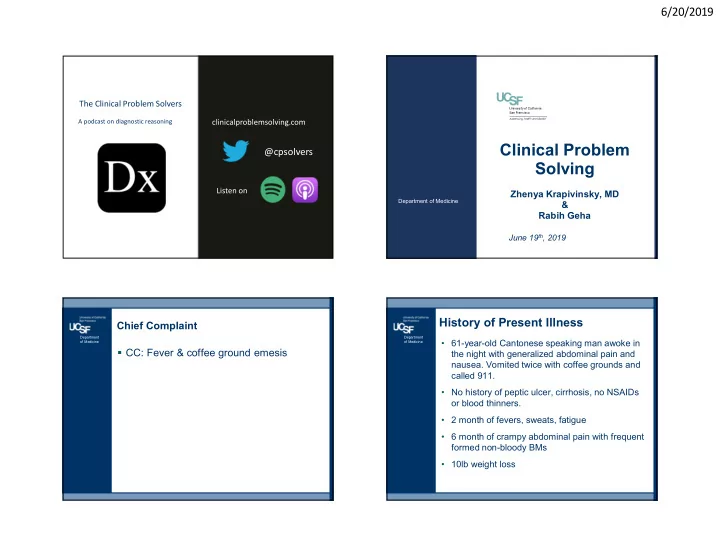

6/20/2019 The Clinical Problem Solvers A podcast on diagnostic reasoning clinicalproblemsolving.com Clinical Problem @cpsolvers Solving Listen on Zhenya Krapivinsky, MD Department of Medicine & Rabih Geha June 19 th , 2019 History of Present Illness Chief Complaint Department Department • 61-year-old Cantonese speaking man awoke in of Medicine of Medicine CC: Fever & coffee ground emesis the night with generalized abdominal pain and nausea. Vomited twice with coffee grounds and called 911. • No history of peptic ulcer, cirrhosis, no NSAIDs or blood thinners. • 2 month of fevers, sweats, fatigue • 6 month of crampy abdominal pain with frequent formed non-bloody BMs • 10lb weight loss 1
6/20/2019 PMH Soc ial History CAD (CABG 10y ago) Postal Officer Department of Medicine HTN Moved to Bay Area from China in1987 HLD Hypothyroidism No recent travel GERD Former smoker (25ppd, quit 15y ago) Medications Clinical Problem Drinks ½ glass whisky/day Aspirin No illicit drug use Atorvastatin Solving Divorced Metoprolol Sexually active with Lisinopril girlfriend for the past year Levothyroxine Father of 3 children & proud June 19 th , 2019 Department of Medicine Omeprazole grandfather of 6 NKDA Stop 1 Family History Father: HTN, DM2 Physical Exam Department of Medicine BP 92/58 | Pulse 112 | Temp 38.8 C | RR 22 |SpO2 98% GEN: frail looking male, no apparent distress HEENT: dry mucous membranes HEART: regular tachycardia, S4, no m/r/g Clinical Problem LUNGS: clear to auscultation bilaterally. no wheezes, rales, Solving or rhonchi ABD: soft, diffusely tender with greatest pain in the LLQ, hypoactive bowel sounds. DRE: brown stool, guaiac +. June 19 th , 2019 Department of Medicine EXT: no LE edema, equal pulses Stop 2 NEURO: unremarkable neurological exam 2
6/20/2019 Labs and Studies Additional Labs and Studies Department Department of Medicine of Medicine Stool studies: 136 98 31 9.4 144 12.3 430 Shigella, salmonella, e.coli: negative 3.6 19 1.5 28 C. diff: negative Ova & parasites: negative LFTs: AST 78 ALT 114 tbili 1.2 AP 68 HBsAg positive INR: 1.2 HBcAg positive Lactate: 3.2 HBV viral Load: 2.5 billion copies per milliliter BCx x2: no growth HCV antibody negative Ucx: no growth CXR: clear lungs Imaging Hospital Course Department Department of Medicine of Medicine CT abdomen Hemoglobin remained stable over the course of three days and there was no recurrence of Mesenteric inflammation with fat stranding, hematemesis. edema and thickening of the bowel wall along the distal sigmoid and rectum. Patient continued to have frequent small non Findings concerning for ischemic colitis. bloody stools. Upper Endoscopy 2 episodes of fevers recorded on day 1 & 2. Erosive esophagitis and mild gastritis without active ulcer or bleed. Biopsies taken for H.pylori. 3
6/20/2019 Discharge Diagnoses & Hospital Course Department of Medicine Ischemic Colitis – Patient was presumed to have ischemic colitis – Treated with bowel rest, IVF, IV Ceftriaxone and Metronidazole Clinical Problem Gastritis & esophagitis Solving – Gastric bx: non-specific inflammation, H .pylori (-) – Initially treated with IV PPI & then oral PPI – No recurrence of hematemesis. June 19 th , 2019 Department of Medicine – Hgb remained stable at 9.3. Stop 3 Discharged after 3 days with outpatient GI & Hepatology follow-up. 2 Months Later…. Review of Systems Department Department of Medicine of Medicine BIBA after a ground level fall on the street Bilateral foot & leg pain that wakes him up from sleep CC: leg weakness that started during the prior Ongoing fevers & sweats hospitalization and continued to worsen post Ongoing abdominal pain, especially after eating discharge. Occasional diarrhea, sometimes with small amounts Has fallen several times over the past 2 months & of blood. has started using a cane. Severe fatigue Due to weakness and difficulty ambulating he had Weight loss missed his GI and Hepatology appointments. Also weakness in his hands, now daughter helps All other ROS are negative him get dressed. 4
6/20/2019 Physical Exam Department BP 160/92 | Pulse 108| Temp 38.5C| RR 22 |SpO2 98% of Medicine GEN: frail looking male with bitemporal wasting, A&O x 3 in no apparent distress HEENT: dry mucous membranes, pale conjunctiva HEART: regular tachycardia, S4, no m/r/g Clinical Problem LUNGS: clear to auscultation bilaterally ABD: soft, diffusely tender. DRE: brown stool, guaiac + Solving EXT: Interosseous muscle wasting in the R hand, calf circumference L< R NEURO: 4/5 wrist extension on the right, 3/5 right finger June 20 th , 2018 Department of Medicine abduction. Left ankle unable to plantar or dorsiflex. Unable Stop 4 to feel light touch on dorsum of left foot, DTR 1+ in left ankle. On walking the patient was observed to have a left foot drop. Otherwise unremarkable neurological exam Labs and Studies Additional Labs Department Department of Medicine of Medicine HIV negative 128 28 98 7.8 126 664 ANA 1:80 18 19 1.3 3.0 dsDNA negative 23.4 ANCA: 1:20 pANCA pattern LFTs: AST 72 ALT 158 tbili 1.4 AP 89 SPEP: slight elevation in the gamma globulin level Albumin: 1.2 without a clear monoclonal band. INR: 1.4 Cryoglobulins: none seen Lactate: 1.9 Complements: mildly elevated C4, normal C3 ESR: 68 CRP: 16.5 CK: 43 (normal) UA: bland Blood gram stain: negative; culture: no growth 5
6/20/2019 Additional Labs and Studies Department of Medicine EMG – Asymmetric, axonal sensory and motor polyneuropathy, primarily affecting the legs but also involving the right arm. Clinical Problem Solving Neurology & Neurosurgery consulted Patient undergoes biopsies of the left peroneal June 20 th , 2018 Department of Medicine nerve. Stop 5 Hospital Course Upper GI Endoscopy Department Department Acute episodes of bright red blood per rectum on of Medicine of Medicine Fresh blood in the stomach and adherent clot in hospital day 3. the fundus over a possible superficial ulcer. – BP 86/54. HR 132 T 39 RR28 O2: 96% on RA – CV: weak peripheral pulses, cold extremities, tachycardia – Abd: tenderness LLQ without guarding. DRE: bright red blood Resuscitated with IVF & 4 units of PRBC EGD: fresh blood in the stomach and adherent clot in the fundus over a possible superficial ulcer (treated endoscopically) 6
6/20/2019 Hospital Course Gastric Dieulafoy's Lesion Department Department of Medicine of Medicine Maroon stool persisted after endoscopy and patient required 6 more units of blood. Repeat endoscopy 2 days later showed a Dieulafoy's lesion in the fundus of stomach with adherent clot. The surrounding mucosa was edematous with submucosal hemorrhage Biopsies were not taken due to high risk of bleeding 36 hours later… Department of Medicine Recurrent hematemesis and hypotension. A diagnostic procedure was performed… Clinical Problem Solving June 19 th , 2019 Department of Medicine Stop 6 7
6/20/2019 What is the most likely diagnosis? CT Angiography Department Department of Medicine of Medicine A. Endocarditis Diffuse irregularity of the B. Syphilis branches of the SMA with 56% C. Tuberculosis multiple narrowing & pseudoaneurysms. D. Polyarteritis Nodosa 38% E. Microangiopathic polyangiitis Fusiform pseudoaneurysm F. SLE in the main splenic artery G. Ehlers-Danlos Syndrome with contrast extravasation from several short gastric H. Atrial Myxoma 6% arteries nodes. 0% 0% 0% 0% 0% Endocarditis Microangiopathic polyangiitis Syphilis Tuberculosis Polyarteritis Nodosa Ehlers-Danlos Syndrome SLE Atrial Myxoma Peroneal nerve biopsy – Active vasculitis with ischemic damage to Department Department the nerve and acute axonal degeneration of Medicine of Medicine Putting it all together: – Medium vessel microaneurysms on angiography – Peroneal nerve vasculitis – Hepatitis B infection Diagnosis: Polyarteritis Nodosa 8
Recommend
More recommend