
SLIDE 1
1
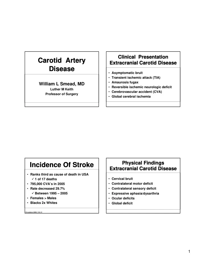
Carotid Artery Disease Carotid Artery Disease
William L Smead, MD
Luther M Keith Professor of Surgery
Incidence Of Stroke Incidence Of Stroke
- Ranks third as cause of death in USA
1 of 17 deaths
- 795,000 CVA’s in 2005
- Rate decreased 29.7%
Between 1995 – 2005
- Females > Males
- Blacks 2x Whites
Circulation 2009; 119: 21
Clinical Presentation Extracranial Carotid Disease Clinical Presentation Extracranial Carotid Disease
- Asymptomatic bruit
- Transient ischemic attack (TIA)
( )
- Amaurosis fugax
- Reversible ischemic neurologic deficit
- Cerebrovascular accident (CVA)
- Global cerebral ischemia
Physical Findings Extracranial Carotid Disease Physical Findings Extracranial Carotid Disease
- Cervical bruit
- Contralateral motor deficit
- Contralateral sensory deficit
- Expressive aphasia/dysarthria
- Ocular deficits
- Global deficit