
SLIDE 1
1
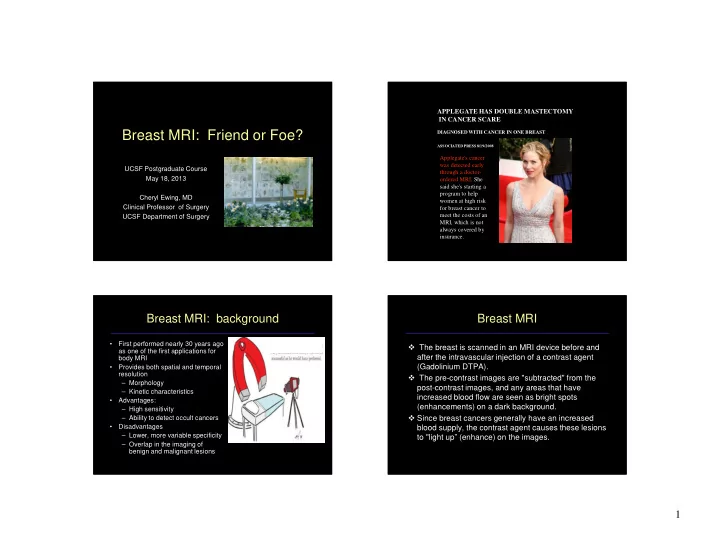
Breast MRI: Friend or Foe?
UCSF Postgraduate Course May 18, 2013 Cheryl Ewing, MD Clinical Professor of Surgery UCSF Department of Surgery APPLEGATE HAS DOUBLE MASTECTOMY IN CANCER SCARE
DIAGNOSED WITH CANCER IN ONE BREAST
- Comments: 0
ASSOCIATED PRESS 8/19/2008
Applegate's cancer was detected early through a doctor-
- rdered MRI. She
said she's starting a program to help women at high risk for breast cancer to meet the costs of an MRI, which is not always covered by insurance.
Breast MRI: background
- First performed nearly 30 years ago
as one of the first applications for body MRI
- Provides both spatial and temporal
resolution – Morphology – Kinetic characteristics
- Advantages:
– High sensitivity – Ability to detect occult cancers
- Disadvantages
– Lower, more variable specificity – Overlap in the imaging of benign and malignant lesions