
SLIDE 1
1
Detection and Treatment of Non- Melanoma Skin Cancers
Toby Maurer, MD
University of California, San Francisco
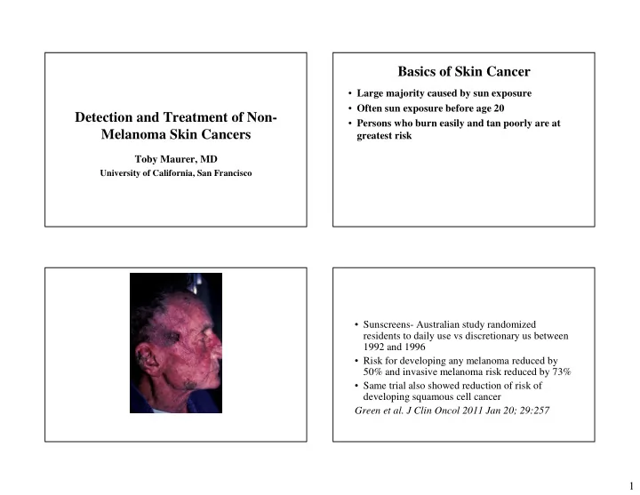
Basics of Skin Cancer
- Large majority caused by sun exposure
- Often sun exposure before age 20
- Persons who burn easily and tan poorly are at
greatest risk
- Sunscreens- Australian study randomized
residents to daily use vs discretionary us between 1992 and 1996
- Risk for developing any melanoma reduced by
50% and invasive melanoma risk reduced by 73%
- Same trial also showed reduction of risk of