
SLIDE 1
5/11/2013 1 A Biomechancial Comparison of an External Fixator to Internal Fixators in an Unstable Pelvic Fracture Model
Erik McDonald, BS; Patrick Horst, MD; Utku Kandemir, MD, Murat Pekmezci, MD University of California San Francisco, San Francisco General Hospital
Background
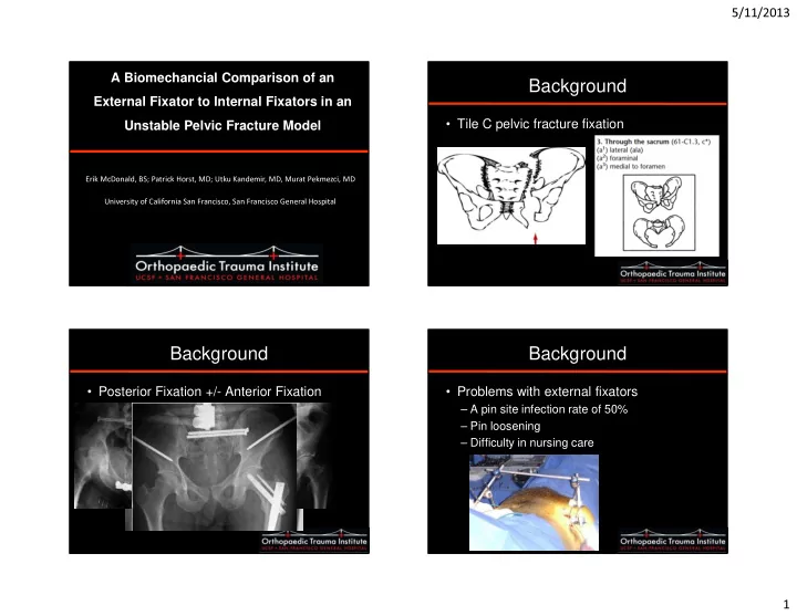
- Tile C pelvic fracture fixation
Background
- Posterior Fixation +/- Anterior Fixation
Background
- Problems with external fixators