SLIDE 1 Page 1/38
An Update on the Genetics, Clinical Presentation and Pathomechanisms of Human
1
Riboflavin Transporter Deficiency
2
Benjamin O’Callaghan1, Annet M Bosch2, Henry Houlden1*
3
1MRC Centre for Neuromuscular Diseases, Department of Neuromuscular Diseases, UCL
4
Queen Square Institute of Neurology and National Hospital for Neurology and Neurosurgery,
5
Queen Square, London WC1N 3BG, UK
6
2Amsterdam UMC, University of Amsterdam, Pediatric Metabolic Diseases, Emma
7
Children's Hospital, Meibergdreef 9, Amsterdam, Netherlands.
8 9
*Corresponding: h.houlden@ucl.ac.uk Tel: 020 7837 3611
10 11
Manuscript word count: 4114
12
Summary Word Count: 174
13
Tables: 1 Figs: 1
14 15
SUMMARY: Riboflavin Transporter Deficiency (RTD) is a rare neurological condition that
16
encompasses the Brown-Vialetto-Van Laere and Fazio-Londe syndromes since the discovery
17
- f pathogenic mutations in the SLC52A2 and SLC52A3 genes that encode human riboflavin
18
transporters RFVT2 and RFVT3. Patients present with a deteriorating progression of
19
peripheral and cranial neuropathy that causes muscle weakness, vision loss, deafness, sensory
20
ataxia and respiratory compromise which when left untreated can be fatal. Considerable
21
progress in the clinical and genetic diagnosis of RTDs has been made in recent years and has
22
permitted the successful lifesaving treatment of many patients with high dose riboflavin
23
supplementation.
24
SLIDE 2
Page 2/38
In this review we first outline the importance of riboflavin and its efficient transmembrane
25
transport in human physiology. Reports on 109 patients with a genetically confirmed
26
diagnosis of RTD are then summarised in order to highlight commonly presenting clinical
27
features and possible differences between patients with pathogenic SLC52A2 (RTD2) or
28
SLC52A3 (RTD3) mutations. Finally, we focus attention on recent work with different
29
models of RTD that have revealed possible pathomechanisms contributing to
30
neurodegeneration in patients.
31 32
Take Home Message: Here we outline the genetics, clinical features, and underlying
33
pathomechanisms of human riboflavin transporter deficiencies (RTDs). Lifesaving treatment
34
with oral riboflavin should be started as soon as a RTD is suspected and continued until the
35
diagnosis has been confirmed or excluded by genetic evaluation.
36 37
COMPLIANCE WITH ETHICS GUIDELINES
38
Author Contributions: Ben O’Callaghan drafted the article. Annet Bosch and Henry
39
Houlden conceived and revised the content.
40
Guarantor: Ben O’Callaghan serves as guarantor for the article.
41
Corresponding Author: Henry Houlden
42
Conflict of Interest: Ben O’Callaghan, Annet Bosch and Henry Houlden declare that they
43
have no conflict of interest.
44
Funding: Ben O’Callaghan is supported by a PhD studentship from the MRC Centre for
45
Neuromuscular Diseases.
46
Ethics Approval: This article does not contain any studies with human or animal subjects
47
performed by any of the authors, and does not require ethics approval.
48
Keywords: SLC52A2, SLC52A3, RFVT, riboflavin, RTD
49
SLIDE 3
Page 3/38
INTRODUCTION
50
Riboflavin belongs to the metabolic B class of vitamins (Vitamin B2) and is the sole
51
precursor for the biologically active cofactors flavin mononucleotide (FMN) and flavin
52
adenine dinucleotide (FAD). During evolution, humans and other higher animals have lost
53
the ability to synthesise riboflavin and instead rely on dietary sources. Emphasising the
54
importance of riboflavin in human physiology and furthermore its efficient absorption and
55
homeostasis are the riboflavin transporter deficiencies (RTDs) (ORPHA 97229
56
https://www.orpha.net/; OMIM 211500, 211530 and 614707) caused by recessive, biallelic
57
mutations in the genes encoding human riboflavin transporters (RFVTs).
58
Essential Role of Riboflavin in Human Physiology
59
Following cellular absorption, riboflavin is rapidly converted into activated flavin cofactors:
60
FMN through riboflavin kinase (RFK: EC 2.7.1.26) mediated phosphorylation of riboflavin,
61
and subsequently FAD by flavin adenine dinucleotide synthetase 1 (FLAD1: EC 2.7.7.2)
62
mediated adenylation of FMN. FMN and FAD are incorporated into 90 different proteins
63
collectively termed the “flavoproteome” (Lienhart et al. 2013), the large majority of which
64
are oxidoreductases localised to the mitochondria that catalyse electron transfer during
65
various redox metabolic reactions including: oxidative decarboxylation of amino acids and
66
glucose, and β-oxidation of fatty acids. Of particular note are a collection of flavoproteins
67
that are crucial for mitochondrial oxidative phosphorylation (OXPHOS) function including:
68
electron-transferring flavoprotein (ETF) and electron-transferring flavoprotein-
69
dehydrogenase (ETFDH: EC 1.5.5.1), which together transfer electrons from various reduced
70
flavin groups to Complex III via Coenzyme Q10; and constituent subunits of Complexes I
71
(NADH Ubiquinone Oxidoreductase Core Subunit V1, NDUFV1: EC 1.6.99.3) and II
72
(Succinate Dehydrogenase Subunit A, SDHA: EC 1.3.5.1).
73
SLIDE 4 Page 4/38
Central to the successful incorporation of flavin cofactors into mitochondrial flavoproteins is
74
the transport of FAD from the cytosol, into the mitochondrial matrix by the mitochondrial
75
FAD transporter (MFT encoded by SLC25A32). Biallelic mutations in SLC25A32 have been
76
associated with riboflavin-responsive exercise intolerance (Schiff et al. 2016) and more
77
recently a severe neuromuscular phenotype (Hellebrekers et al. 2017), highlighting the
78
subcellular importance of flavin availability within mitochondria in particular. For further
79
discussion on the mitochondrial FAD transporter, readers are referred to an accompanying
80
review in this issue that addresses disorders of riboflavin metabolism (Balasubramaniam et
81
82
Other important roles of flavoproteins include: the activation of other B class vitamins, redox
83
homeostasis, transcriptional regulation through enzymatic chromatin modifications, caspase
84
independent apoptosis and cytoskeletal reorganisation (Lienhart et al. 2013; Barile et al.
85
2016).
86
Considering the importance of flavins in metabolically active cells it is unsurprising that
87
inadequate supply of riboflavin has been implicated in diseases of energy demanding tissues,
88
particularly the nervous system.
89
Human Riboflavin Transporters
90
In order to maintain a sufficient supply of flavins to cells throughout the body, humans and
91
- ther higher animals have established an effective carrier-mediated system to transport
92
riboflavin across plasma membranes. Three human RFVT homologues have been identified:
93
RFVT1-3 encoded by genes SLC52A1-3 respectively (note RFVT2 and RFVT3 were
94
designated RFT3 and RFT2 respectively in previous nomenclature) (Yonezawa et al. 2008;
95
Yamamoto et al. 2009; Yao et al. 2010; Yonezawa and Inui 2013). RFVT1 and RFVT2
96
display 87 % amino acid sequence identity, whereas RFVT3 only exhibits 44 % and 45 %
97
SLIDE 5 Page 5/38
amino acid sequence identity with RFVT1 and RFVT2 respectively (ClustalW:
98
http://www.clustal.org/omega/ ).
99
Transmembrane Topology
100
Some confusion surrounding the transmembrane (TM) topology of RFVTs is present in the
101
- literature. Based on initial in silico predictions, RFVT1 and RFVT2 were predicted to have
102
10 TM domains (Yonezawa et al. 2008; Yao et al. 2010) whereas RFVT3 was predicted to
103
have 11 TM domains (Yonezawa and Inui 2013). In silico predictions made using other
104
membrane topology algorithms predict all three RFVTs to have 11 TM domains however
105
(Yamamoto et al. 2009; Udhayabanu et al. 2016; Colon-Moran et al. 2017), and this is
106
supported by immunostaining of hemagglutinin (HA) tagged RFVT1 constructs that indicate
107
an intracellular N-terminus and extracellular C-terminus (Mattiuzzo et al. 2007). Knowing
108
the correct RFVT topology might be important for correlating disease causing mutation sites
109
with differences in phenotypical presentations and/or responsiveness to therapeutic
110
interventions.
111
Tissue Distribution
112
mRNA expression of the three different RFVT genes in human tissues has been assessed
113
(Yao et al. 2010) and is largely in accordance with more recent gene expression data from the
114
GTEx V7 dataset (https://gtexportal.org/). SLC52A1 is mainly expressed in the placenta and
115
- intestine. SLC52A2 is rather ubiquitously expressed but is particularly abundant in nervous
116
- tissues. SLC52A3 is most highly expressed in testis but also intestine and prostate. These
117
different but overlapping expression profiles might explain the vulnerability of certain tissues
118
to mutations in one or more of the SLC52A genes.
119 120 121
SLIDE 6 Page 6/38
RIBOFLAVIN TRANSPORTER DEFICIENCIES (RTDs)
122
Brown-Vialetto-Van Laere (BVVL) and Fazio-Londe (FL) are two phenotypically
123
continuous syndromes presenting with a progressive sensorimotor and cranial neuropathy.
124
Both share a core phenotype of: bulbar palsy (e.g. dysphagia, dysphonia, tongue atrophy),
125
axial and distal muscle weakness, optic atrophy, sensory ataxia and respiratory compromise
126
due to diaphragm paralysis (Bosch et al. 2011; Horvath 2012; Manole and Houlden 2015;
127
Jaeger and Bosch 2016). Sensorineural deafness is present in BVVL only. Since 2010
128
biallelic mutations in the human riboflavin transporter genes SLC52A3 (previously C20orf54)
129
and SLC52A2 have been demonstrated to be the cause of the BVVL and FL syndromes which
130
were renamed to Riboflavin Transporter Deficiencies (RTDs) (Green et al. 2010; Johnson et
131
- al. 2010, 2012, Bosch et al. 2011, 2012; Foley et al. 2014; Manole and Houlden 2015). RTD2
132
and RTD3 refer to disorders caused by SLC52A2 and SLC52A3 mutations respectively
133
(Tables S2 and S3).
134
Transient Riboflavin Deficiency
135
Although pathogenic mutations in SLC52A1 have not been described in patients with a
136
typical RTD phenotype, there have been two reports of transient riboflavin deficiency
137
- ccurring in the newborn children of mothers harbouring one heterozygous SLC52A1
138
mutation (OMIM 615026), in one case in combination with a riboflavin deficiency due to
139
deficient maternal intake (Table S1) (Ho et al. 2011; Mosegaard et al. 2017). In both cases the
140
children but not the mothers showed clinical symptoms of riboflavin deficiency after birth
141
that had subsided by two years of age. Whilst SLC52A1 is expressed in both the human small
142
intestine and placenta, the transient nature of the clinical presentation suggests that these
143
cases were caused by placental haploinsufficiency, and associated impairment in the transport
144
- f riboflavin from the mother to the fetus.
145 146
SLIDE 7 Page 7/38
Genetically Diagnosed Cases of Riboflavin Transporter Deficiency
147
An article in this journal three years ago (Jaeger and Bosch 2016) summarised reports of 70
148
genetically confirmed RTD patients that had been published at that time (Green et al. 2010;
149
Johnson et al. 2010, 2012; Bosch et al. 2011; Anand et al. 2012; Koy et al. 2012; Dezfouli et
150
- al. 2012; Haack et al. 2012; Ciccolella et al. 2012, 2013; Spagnoli et al. 2014; Foley et al.
151
2014; Bandettini Di Poggio et al. 2014; Srour et al. 2014; Cosgrove et al. 2015; Horoz et al.
152
2016; Menezes et al. 2016a; Davis et al. 2016).
153
There have since been a further 10 publications reporting on 23 newly diagnosed RTD2 cases
154
(Petrovski et al. 2015; Menezes et al. 2016b; Guissart et al. 2016; Allison et al. 2017; Manole
155
et al. 2017; Woodcock et al. 2017; Çıralı et al. 2017; Babanejad et al. 2018; Nimmo et al.
156
2018; Set et al. 2018), and 12 reporting on 27 newly diagnosed RTD3 cases (van der Kooi et
157
- al. 2016; Manole et al. 2017; Thulasi et al. 2017; Bashford et al. 2017; Chaya et al. 2017;
158
Woodcock et al. 2017; Hossain et al. 2017; Kurkina et al. 2017; Khadilkar et al. 2017;
159
Nimmo et al. 2018; Camargos et al. 2018; Gowda et al. 2018). A patient harbouring a
160
heterozygous pathogenic mutation in SLC52A3 and heterozygous SLC52A2 variant of
161
unknown significance has been described (Allison et al. 2017), which will be considered as a
162
RTD3 case here. The possibility that both heterozygous mutations within the two different
163
riboflavin genes are synergistically disrupting the same metabolic pathway to a pathogenic
164
level cannot be excluded however. Finally, a patient with homozygous mutations in both
165
SLC52A2 and SLC52A3 (Udhayabanu et al. 2016) has also been described (RTD2/3). In total,
166
various degrees of information are available on 109 patients (52 RTD2, 56 RTD3 and 1
167
RTD2/3) with 71 different SLC52A mutations (24 SLC52A2, 47 SLC52A3) (Table 1).
168
SLC52A2 and SLC52A3 Pathogenic Variants
169
Pathogenic variants in SLC52A2 and SLC52A3 are distributed throughout all coding exons
170
(Ex2-5) and include nonsense and missense mutations affecting RFVT amino acid residues
171
SLIDE 8 Page 8/38
constituting: transmembrane domains, intracellular loops, extracellular loops and C-terminus
172
(Tables S2 and S3). Single nucleotide substitutions within intron-exon boundaries have also
173
been identified in SLC52A2 and SLC52A3 that likely cause splicing defects (Bosch et al.
174
2011; Manole et al. 2017; Çıralı et al. 2017). Single/double nucleotide insertions/deletions
175
causing frameshift mutations have been identified in SLC52A3 (Green et al. 2010; Bandettini
176
di Poggio et al. 2013; Manole et al. 2017), in addition to a more recently described in-frame
177
insertion of 60 nucleotides (20 amino acid peptide) (Camargos et al. 2018).
178
Using heterologous expression systems the impact of different pathogenic SLC52A2/3
179
mutations on RFVT2/3 function has been assessed in vitro (Nabokina et al. 2012; Haack et al.
180
2012; Foley et al. 2014; Subramanian et al. 2015; Petrovski et al. 2015; Udhayabanu et al.
181
2016). In most cases the disease causing mutation reduces RFVT cell surface expression
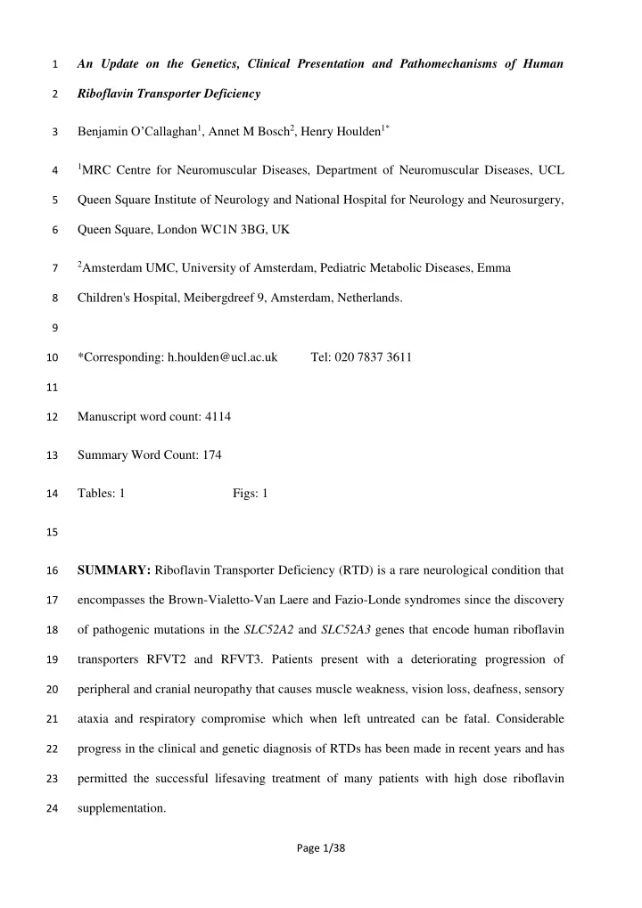
182
which when assessed appears to be due to retainment in the endoplasmic reticulum (ER),
183
indicative of protein misfolding and/or trafficking defect. In some instances riboflavin
184
transport is impaired but with an apparently normal cell surface expression. Of the 15 mutant
185
RFVTs assessed only one (SLC52A3 Genbank NM_033409.3 c.1048T>A; RFVT3
186
p.Leu350Met) has been shown to be functionally normal (Nabokina et al. 2012). Evidence for
187
a reduction in mRNA stability has also been shown for a SLC52A2 single nucleotide
188
substitution (Ciccolella et al. 2013). Finally, impaired riboflavin uptake has been described in
189
fibroblasts from patients harbouring compound heterozygous SLC52A2 mutations (Ciccolella
190
et al. 2013; Manole et al. 2017).
191
Clinical Differentiation of RTD2 and RTD3
192
Disease Onset
193
The large majority of patients with either RTD2 or RTD3 present early in life but until now
194
- nly in RTD3 has a late onset (as late as the third decade) been reported (Bashford et al.
195
2017; Camargos et al. 2018). Late onset RTD (>10y) might therefore be more suggestive of a
196
SLIDE 9 Page 9/38
SLC52A3 mutation. Hearing loss and muscle weakness are among the most common
197
presenting symptoms at onset of both RTD2 and RTD3. Abnormal gait and/or ataxia is often
198
a presenting feature of RTD2 but rarely RTD3. By contrast RTD3 commonly presents with
199
bulbar symptoms, whereas in RTD2 these are generally observed later in the disease course.
200
Other symptoms regularly described upon RTD onset include hypotonia, facial weakness and
201
respiratory dysfunction due to diaphragmatic paralysis as well as muscle weakness.
202
Common Symptoms
203
Whilst hearing loss as a consequence of cranial nerve VIII degeneration is a presenting
204
symptom of many patients, others develop sensorineural hearing loss later in the disease
205
course, and this remains the most commonly observed clinical feature of RTD2 and RTD3.
206
Bulbar symptoms such as dysphagia and dysarthria are present in most patients and a large
207
number display feeding difficulties as a result of dysphagia that in many instances
208
necessitates a nasogastric tube or gastrostomy feeding device. Artificial respiratory devices
209
are also often required, with respiratory symptoms due to neurogenic diaphragm paralysis
210
being very common. Weakness and hypotonia of both limb and axial muscles was prevalent
211
and commonly associated with neurogenic muscular atrophy, particularly of distal muscles.
212
Facial weakness caused by cranial nerve VII (facial nerve) degeneration was common in
213
RTD3 but rarely seen in RTD2. Abnormal gait and/or ataxia remains a distinguishing feature
214
- f RTD2, with RTD3 patients rarely showing signs later during the disease course. SLC52A2
215
mutations have recently been associated with spinocerebellar ataxia with blindness and
216
deafness type 2 (SCABD2) (Guissart et al. 2016; Babanejad et al. 2018). Finally, vision loss
217
caused by cranial nerve II (optic nerve) atrophy was observed in numerous RTD3 cases but
218
appears to be a much more prevalent feature of RTD2.
219 220 221
SLIDE 10
Page 10/38
Neurodiagnostic Tests
222
Neurophysiological studies are suggestive of peripheral neuropathy in the large majority of
223
RTD patients tested but normal results are also observed, particularly in RTD3. Motor and
224
sensory nerve conduction studies are indicative of an axonal rather than demyelinating
225
neuropathic phenotype, with signs of anterior horn dysfunction and chronic denervation in
226
most RTD cases. Slightly slowed sensorimotor conduction velocities suggestive of
227
demyelination have been described in a minority of RTD2 cases (Guissart et al. 2016; Allison
228
et al. 2017), and a single RTD3 patient (Bandettini di Poggio et al. 2013; Bandettini Di
229
Poggio et al. 2014) however.
230
In the large majority of RTD cases brain magnetic resonance imaging (MRI) is unremarkable.
231
Abnormal MRI observations rarely described in RTD2 brain include: mild atrophy of the
232
cerebellar vermis (Guissart et al. 2016), optic nerve abnormalities (Woodcock et al. 2017; Set
233
et al. 2018) and thinning/shortening of the corpus callosum (Srour et al. 2014; Set et al.
234
2018). Cerebellar abnormalities described in RTD3 brain MRI include: hyperintense T2-
235
weighted signals within cerebellar peduncles (Koy et al. 2012; Bandettini Di Poggio et al.
236
2014), and volume loss of peduncles and vermis over an 8 year period (Bandettini Di Poggio
237
et al. 2014). Intense T2-weighted signals have also been noted in cortical, subcortical (basal
238
ganglia and internal capsule) and brainstem (vestibular nuclei and central tegmental tract)
239
regions of some RTD3 patients (Koy et al. 2012; Spagnoli et al. 2014; Hossain et al. 2017;
240
Nimmo et al. 2018). Spinal MRI has been conducted much less frequently, but abnormal T2-
241
weighted intensities have been described in ventral nerve roots and dorsal regions of the
242
spinal cord (Koy et al. 2012; Spagnoli et al. 2014; Davis et al. 2016; Woodcock et al. 2017;
243
Khadilkar et al. 2017) in accordance with the sensorimotor phenotype of RTD.
244 245 246
SLIDE 11 Page 11/38
Neuropathology
247
Assessment of sural nerve biopsies from RTD2 (Haack et al. 2012; Foley et al. 2014; Srour et
248
- al. 2014) and RTD3 (Johnson et al. 2010; Chaya et al. 2017) patients show evidence for
249
axonal neuropathy and degeneration which preferentially affects large calibre myelinated
250
axons (Foley et al. 2014; Srour et al. 2014; Chaya et al. 2017), in accordance with the sensory
251
impairments observed in these patients.
252
Recent neuropathological observations described in the central nervous system of two RTD3
253
patients are also reflective of the RTD clinical phenotype (Manole et al. 2017). In line with
254
the bulbar symptoms that are commonly observed, nuclei and tracts of cranial nerves IX, X
255
and XII showed marked neuronal loss and gliosis. Loss of neurons was also observed in the
256
nuclei of cranial nerves III and IV in accordance with eye movement impairments observed
257
in these patients. The nuclei of cranial nerve VIII and tracts of cranial nerve II showed
258
evidence of degeneration, underscoring the clinical presentation of sensorineural deafness
259
and vision loss respectively. Gliosis and neuronal loss was also evident in midbrain (medial
260
lemniscus, central tegmental tract) brainstem (pons, medulla), cerebellum (white matter
261
structures including cerebellar peduncles, cerebellar nuclei) and spinal cord (anterior horn,
262
spinothalamic tracts, spinocerebellar tracts), fitting with MRI observations that have been
263
made in some RTD3 patients (see above). Of particular interest was the presence of
264
symmetrical lesions in the brainstem of both patients that showed demyelination and
265
macrophage infiltration but with relative sparing of the neurons. The authors highlighted the
266
similarities of these lesions to neuropathological observations made in mitochondrial disease
267
patients.
268
Biochemical Tests
269
An increase in plasma acylcarnitines is indicative of an impairment in the metabolism of fatty
270
acids by mitochondrial β-oxidation and is a characteristic observation of the multiple acyl-
271
SLIDE 12 Page 12/38
CoA dehydrogenation defect (MADD) syndromes caused by mutations in ETF (encoded by
272
ETFA and ETFB) or ETFDH (encoded by ETFDH) flavoproteins (OMIM 231680).
273
Identification of a MADD-like acylcarnitine profile in BVVL patients without ETFA, ETFB
274
- r ETFDH mutations led to a hypothesis of impaired riboflavin absorption and was key to the
275
initial identification of BVVL as a RTD (Bosch et al. 2011). However, nearly half of the
276
RTD cases described since show normal acylcarnitine profiles on diagnosis and thus it cannot
277
be used to exclude a RTD diagnosis.
278
Urine organic acid analysis has been reported less frequently, and in half of RTD cases
279
results are normal. Ethylmalonic aciduria suggestive of impairments in fatty-acid, methionine
280
and/or isoleucine oxidation is the most common abnormality noted (4/10 RTD2, 5/12 RTD3).
281
Flavoproteins constitute important steps in the metabolic pathways responsible for branched-
282
chain, lysine and tryptophan amino acid catabolism (Barile et al. 2016) and elevations in
283
acylglycines associated with impairments of such pathways have also been described.
284
Assessment of plasma flavin status necessitates mass spectrometry analysis and is not
285
routinely done in the clinical setting. In the small number of patients assessed, plasma flavin
286
levels are generally within the normal range but low levels have been reported in both RTD2
287
(Srour et al. 2014) and RTD3 (Bosch et al. 2011). Following high dose riboflavin treatment,
288
increases in plasma flavin levels are observed in both RTD2 and RTD3 cases (Bosch et al.
289
2011; Haack et al. 2012; Foley et al. 2014), highlighting the partial redundancy of RFVT
290
homologues in intestinal absorption. Nevertheless, with many patients presenting with normal
291
flavins at diagnosis, plasma flavin status cannot be used as a tool to exclude a RTD diagnosis.
292
Measurements of the erythrocyte glutathione reductase activity coefficient (EGRAC) are
293
representative of flavin status and more routinely done in the clinic. An abnormal EGRAC
294
measurement without acylcarnitine abnormalities has been reported in a single RTD3 case,
295
which normalised following riboflavin supplementation (Chaya et al. 2017).
296
SLIDE 13 Page 13/38
Genetic Diagnostic Strategy
297
Whilst there does appear to be differences in the commonest clinical signs linked with RTD2
298
- r RTD3, there is no observation that can definitively distinguish between the two. It is
299
therefore recommended that genetic analysis of SLC52A2 and SLC52A3 is performed
300
simultaneously rather than sequentially in suspected RTD cases (Manole and Houlden 2015).
301
Even though mutations in SLC52A1 are yet to be associated with a typical RTD phenotype, it
302
remains a viable candidate that should also be considered. Whole or focused exome analysis
303
using next generation sequencing (NGS) technology might then be performed, with a filtering
304
strategy targeting genes associated with: similar clinical phenotypes (e.g. amyotrophic lateral
305
sclerosis, OMIM 105400; Joubert syndrome, OMIM 213300; Nathalie syndrome, OMIM
306
255990; Madras motor neuron disease, ORPHA 137867; MADD, OMIM 231680), riboflavin
307
metabolism, the flavoproteome and/or mitochondrial metabolism.
308
High Dose Riboflavin Therapy
309
Identification of causative SLC52A mutations in these debilitating disorders has not only
310
advanced their genetic diagnoses but also highlighted high dose oral riboflavin
311
supplementation as an effective therapeutic intervention. Excess riboflavin is excreted in the
312
urine and toxicity has not been reported, making riboflavin therapy a safe intervention. Over
313
70 % of patients demonstrate improvements in muscle strength, motor abilities, respiratory
314
function and/or cranial nerve deficits, with some patients no longer requiring ventilatory
315
- support. No deaths have been reported in riboflavin treated patients, whilst over half of
316
untreated patients reported have died (Jaeger and Bosch 2016).
317
Effective doses which have been used vary between 10-80 mg/kg/day, whilst doses below 10
318
mg/kg/day are reported to be ineffective (personal communications). Doses as high as 80
319
mg/kg body weight per day (Chaya et al. 2017; Forman et al. 2018) have been tolerated with
320
SLIDE 14 Page 14/38
minimal side effects, although gastrointestinal side effects are rarely noted (Bosch et al. 2011;
321
Foley et al. 2014; Woodcock et al. 2017; Nimmo et al. 2018).
322
Responses to high dose riboflavin are similarly observed in the majority of RTD2 and RTD3
323
cases (i.e. genotype is not predictive of treatment response). Clinical improvement following
324
riboflavin treatment is observed for the majority of RTD patients (19/30 RTD2, 20/23 RTD3)
325
with the remaining patients showing stabilisation of the current disease state (10/30 RTD2,
326
1/23). In only two RTD3 patients has no beneficial response to riboflavin supplementation
327
been reported. In one of these cases treatment was not started until 29 years after disease
328
- nset (Davis et al. 2016), at which point irreversible neurodegenerative changes will have
329
- ccurred. In the second non-responsive case, treatment was discontinued after 1 week (Koy et
330
- al. 2012) which might have preceded a latent response, as clinical improvement is frequently
331
not observed for months following the beginning of treatment. For example patient 2 reported
332
by (Nimmo et al. 2018) was started on 80 mg/kg/day riboflavin at 8 months of age but his
333
ventilator dependency had not improved by 10 months of age and for this reason a
334
tracheostomy was performed. However, with continued riboflavin treatment clinical
335
improvement was observed, and by the age of 14 months he was able to maintain
336
spontaneous respiration.
337
Generally the most positive responses are reported in patients that receive riboflavin
338
supplementation shortly after disease onset (Foley et al. 2014). Of note, a newly born sibling
339
- f an RTD3 patient harbouring the same pathogenic mutations has been administered
340
riboflavin since birth and remains asymptomatic after 1 year (Horoz et al. 2016), whilst
341
patient 2 from the first report of RTD3 who was symptomatic and treated from 3 months of
342
age (Bosch et al. 2011) is now still asymptomatic at 8 years of age.
343
For these reasons it is recommended that riboflavin is administered immediately upon
344
suspected RTD in order to prevent irreversible neurological changes, and continued until an
345
SLIDE 15 Page 15/38
alternate unrelated cause of disease has been identified. Esterified derivatives of riboflavin
346
are less reliant on RFVTs for cellular absorption and might therefore represent a strategy for
347
future RTD therapeutics with improved bioavailability (Manole et al. 2017).
348 349
RECENT INSIGHTS INTO RTD PATHOMECHANISMS
350
Whilst there has been great advancement in RTD diagnosis and treatment, much less progress
351
has been made in determining the pathomechanisms that lead to cranial and peripheral nerve
352
- degeneration. Flavins are important to the function of cells throughout the whole body, yet
353
neurons appear to be especially vulnerable to riboflavin depletion. Recent work has started to
354
unravel possible downstream consequences of RFVT dysfunction that might lead to
355
neurodegeneration (Figure 1).
356
In a study by (Rizzo et al. 2017), human induced pluripotent stem cell (hIPSC) lines were
357
established from a RTD2 and RTD3 patient and differentiated into motor neurons. RTD
358
motor neurons displayed an increase in neurofilament heavy chain (NFH) expression and its
359
aggregation in inclusions, something previously characterised as an early event leading to
360
motor neuron degeneration in amyotrophic lateral sclerosis (ALS) (Chen et al. 2014). An
361
associated reduction in axonal length was also observed, however in a more recent study by
362
(Manole et al. 2017) no such cytoskeletal abnormalities were described in the motor axons of
363
Drosophila with knockdown of the Drosophila homologue of SLC52A (drift).
364
The phenotypic overlap of RTD with primary mitochondrial diseases and important role of
365
flavins in mitochondrial function, might point towards mitochondrial dysfunction as an
366
important pathomechanism contributing to neurodegeneration in RTD. In accordance,
367
mitochondria within neurons of drift knockdown Drosophila are structurally abnormal, show
368
reduced activity of OXPHOS complexes I and II and more depolarised mitochondrial
369
membrane potential (Manole et al. 2017). Such abnormalities are also seen in RTD2
370
SLIDE 16
Page 16/38
fibroblasts (Manole et al. 2017) and RTD muscle biopsies (Foley et al. 2014; Chaya et al.
371
2017; Nimmo et al. 2018). OXPHOS activity is normal in hIPSC-derived RTD motor
372
neurons, but impairments in mitochondrial fusion and autophagy (mitophagy) were seen
373
(Rizzo et al. 2017), both of which are important for maintaining a healthy mitochondrial
374
network in post mitotic cells.
375
Neurons are among the most energy demanding cells of the body making them particularly
376
sensitive to impairments in cellular metabolic processes. Increases in the release of
377
mitochondrial derived reactive oxygen species (ROS) have also been implicated as
378
pathomechanisms contributing to neuronal death. Mitochondrial dysfunction and concurrent
379
impairments in their clearance might therefore be contributing to the specific vulnerability of
380
neurons in RTD patients, and represent an additional pathomechanism that is shared with
381
many other neurodegenerative conditions including primary mitochondrial diseases and ALS
382
(Golpich et al. 2017).
383 384
CONCLUSION
385
The RTDs are an excellent example of how the genetic diagnosis of an inborn error of
386
metabolism can translate an effective rational based therapy back in to the clinic. Although
387
clinical improvements upon riboflavin supplementation are observed in many patients, some
388
cases only show a stabilisation of the current disease state indicating quick intervention with
389
riboflavin supplementation is important to avoid irreversible damage from occurring.
390
Therefore, start of oral riboflavin supplementation upon suspicion of RTD diagnosis without
391
awaiting test results is of utmost importance and lifesaving. Positive clinical responses to
392
riboflavin supplementation might occur with some latency and for this reason riboflavin
393
therapy should be continued in all suspected or genetically diagnosed RTD cases, even if no
394
apparent clinical improvement has initially occurred. In the foreseeable future newborn
395
SLIDE 17 Page 17/38
screening of SLC52A1-3 might ensure riboflavin therapy is administered prior to the
396
presentation of symptoms. Whilst biochemical screening parameters might in some instances
397
be suggestive of RTD, diagnosis can only be made by genetic analysis. Genetic analysis of
398
SLC52A1-3 should therefore be the basis for such newborn screening tests. Understanding the
399
pathomechanisms contributing to irreversible neuronal damage caused by riboflavin
400
depletion might reveal additional targets for novel therapeutic intervention in patients which
401
receive a delayed diagnosis.
402 403
REFERENCES
404
Allison T, Roncero I, Forsyth R, et al (2017) Brown-Vialetto-Van Laere Syndrome as a
405
Mimic of Neuroimmune Disorders: 3 Cases from the Clinic and Review of the
406
- Literature. J Child Neurol 32:528–532. doi: 10.1177/0883073816689517
407
Anand G, Hasan N, Jayapal S, et al (2012) Early use of high-dose riboflavin in a case of
408
Brown-Vialetto-Van Laere syndrome. Dev Med Child Neurol 54:187–9. doi:
409
10.1111/j.1469-8749.2011.04142.x
410
Babanejad M, Adeli OA, Nikzat N, et al (2018) SLC52A2 mutations cause SCABD2
411
phenotype: A second report. Int J Pediatr Otorhinolaryngol 104:195–199. doi:
412
10.1016/J.IJPORL.2017.11.014
413
Balasubramaniam, S. , Christodoulou, J. and Rahman, S. Disorders of Riboflavin
414
- Metabolism. J Inherit Metab Dis.. Accepted Author Manuscript. 2019.
415
doi:10.0.3.234/jimd.12058
416
Bandettini di Poggio M, Gagliardi S, Pardini M, et al (2013) A novel compound
417
heterozygous mutation of C20orf54 gene associated with Brown-Vialetto-Van Laere
418
syndrome in an Italian family. Eur J Neurol 20:e94–e95. doi: 10.1111/ene.12163
419
SLIDE 18 Page 18/38
Bandettini Di Poggio M, Monti Bragadin M, Reni L, et al (2014) Brown-Vialetto-Van Laere
420
syndrome: Clinical and neuroradiological findings of a genetically proven patient.
421
Amyotroph Lateral Scler Front Degener 15:141–144. doi:
422
10.3109/21678421.2013.837931
423
Barile M, Giancaspero TA, Leone P, et al (2016) Riboflavin transport and metabolism in
424
- humans. J Inherit Metab Dis 39:545–557. doi: 10.1007/s10545-016-9950-0
425
Bashford JA, Chowdhury FA, Shaw CE (2017) Remarkable motor recovery after riboflavin
426
therapy in adult-onset Brown-Vialetto-Van Laere syndrome. Pract Neurol 17:53–56.
427
doi: 10.1136/practneurol-2016-001488
428
Bosch AM, Abeling NGGM, Ijlst L, et al (2011) Brown-Vialetto-Van Laere and Fazio Londe
429
syndrome is associated with a riboflavin transporter defect mimicking mild MADD: a
430
new inborn error of metabolism with potential treatment. J Inherit Metab Dis 34:159–64.
431
doi: 10.1007/s10545-010-9242-z
432
Bosch AM, Stroek K, Abeling NG, et al (2012) The Brown-Vialetto-Van Laere and Fazio
433
Londe syndrome revisited: natural history, genetics, treatment and future perspectives.
434
Orphanet J Rare Dis 7:83. doi: 10.1186/1750-1172-7-83
435
Camargos S, Guerreiro R, Bras J, Mageste LS (2018) Late-onset and acute presentation of
436
Brown-Vialetto-Van Laere syndrome in a Brazilian family. Neurol Genet 4:e215. doi:
437
10.1212/NXG.0000000000000215
438
Chaya S, Zampoli M, Gray D, et al (2017) The First Case of Riboflavin Transporter
439
Deficiency in sub-Saharan Africa. Semin Pediatr Neurol. doi:
440
10.1016/J.SPEN.2017.03.002
441
Chen H, Qian K, Du Z, et al (2014) Modeling ALS with iPSCs Reveals that Mutant SOD1
442
Misregulates Neurofilament Balance in Motor Neurons. Cell Stem Cell 14:796–809. doi:
443
SLIDE 19 Page 19/38
10.1016/J.STEM.2014.02.004
444
Ciccolella M, Catteruccia M, Benedetti S, et al (2012) Brown–Vialetto–van Laere and Fazio–
445
Londe overlap syndromes: A clinical, biochemical and genetic study. Neuromuscul
446
Disord 22:1075–1082. doi: 10.1016/J.NMD.2012.05.007
447
Ciccolella M, Corti S, Catteruccia M, et al (2013) Riboflavin transporter 3 involvement in
448
infantile Brown-Vialetto-Van Laere disease: two novel mutations. J Med Genet 50:104–
449
- 7. doi: 10.1136/jmedgenet-2012-101204
450
Çıralı C, Ergin H, Özdemir ÖM, et al (2017) P385 Hypotonic infant with riboflavin
451
transporter deficiency due to slc52a2 mutations. In: Posters. BMJ Publishing Group Ltd
452
and Royal College of Paediatrics and Child Health, p A181.3-A182
453
Colon-Moran W, Argaw T, Wilson CA (2017) Three cysteine residues of SLC52A1, a
454
receptor for the porcine endogenous retrovirus-A (PERV-A), play a critical role in cell
455
surface expression and infectivity. Virology 507:140–150. doi:
456
10.1016/J.VIROL.2017.04.019
457
Cosgrove J, Datta S, Busby M (2015) Adult onset Brown–Vialetto–Van Laere syndrome with
458
- psoclonus and a novel heterozygous mutation: A case report. Clin Neurol Neurosurg
459
128:1–3. doi: 10.1016/J.CLINEURO.2014.10.016
460
Davis A, Josifova D, Lloyd-Owen S, et al (2016) Brown-Vialetto-Van Laere syndrome: a 28-
461
year follow-up. J Neurol Neurosurg Psychiatry 87:681–2. doi: 10.1136/jnnp-2014-
462
310088
463
Dezfouli MA, Yadegari S, Nafissi S, Elahi E (2012) Four novel C20orf54 mutations
464
identified in Brown-Vialetto-Van Laere syndrome patients. J Hum Genet 57:613–7. doi:
465
10.1038/jhg.2012.70
466
Foley AR, Menezes MP, Pandraud A, et al (2014) Treatable childhood neuronopathy caused
467
SLIDE 20 Page 20/38
by mutations in riboflavin transporter RFVT2. Brain 137:44–56. doi:
468
10.1093/brain/awt315
469
Forman EB, Foley AR, King MD (2018) Dramatic improvement of a rare syndrome with
470
high dose riboflavin treatment. Pediatr Neurol. doi:
471
10.1016/J.PEDIATRNEUROL.2018.05.005
472
Golpich M, Amini E, Mohamed Z, et al (2017) Mitochondrial Dysfunction and Biogenesis in
473
Neurodegenerative diseases: Pathogenesis and Treatment. CNS Neurosci Ther 23:5–22.
474
doi: 10.1111/cns.12655
475
Gowda VK, Udhayabanu T, Varalakshmi P, et al (2018) Fazio-Londe syndrome in siblings
476
from India with different phenotypes. Brain Dev. doi:
477
10.1016/J.BRAINDEV.2018.02.010
478
Green P, Wiseman M, Crow YJ, et al (2010) Brown-Vialetto-Van Laere syndrome, a ponto-
479
bulbar palsy with deafness, is caused by mutations in c20orf54. Am J Hum Genet
480
86:485–9. doi: 10.1016/j.ajhg.2010.02.006
481
Guissart C, Drouot N, Oncel I, et al (2016) Genes for spinocerebellar ataxia with blindness
482
and deafness (SCABD/SCAR3, MIM# 271250 and SCABD2). Eur J Hum Genet
483
24:1154–1159. doi: 10.1038/ejhg.2015.259
484
Haack TB, Makowski C, Yao Y, et al (2012) Impaired riboflavin transport due to missense
485
mutations in SLC52A2 causes Brown-Vialetto-Van Laere syndrome. J Inherit Metab
486
Dis 35:943–8. doi: 10.1007/s10545-012-9513-y
487
Hellebrekers DMEI, Sallevelt SCEH, Theunissen TEJ, et al (2017) Novel SLC25A32
488
mutation in a patient with a severe neuromuscular phenotype. Eur J Hum Genet 25:886–
489
- 888. doi: 10.1038/ejhg.2017.62
490
Ho G, Yonezawa A, Masuda S, et al (2011) Maternal riboflavin deficiency, resulting in
491
SLIDE 21
Page 21/38
transient neonatal-onset glutaric aciduria Type 2, is caused by a microdeletion in the
492
riboflavin transporter gene GPR172B. Hum Mutat 32:E1976–E1984. doi:
493
10.1002/humu.21399
494
Horoz OO, Mungan NO, Yildizdas D, et al (2016) Brown-Vialetto-Van Laere syndrome: two
495
siblings with a new mutation and dramatic therapeutic effect of high-dose riboflavin. J
496
Pediatr Endocrinol Metab 29:227–231. doi: 10.1515/jpem-2015-0198
497
Horvath R (2012) Update on clinical aspects and treatment of selected vitamin-responsive
498
disorders II (riboflavin and CoQ10). J Inherit Metab Dis 35:679–687. doi:
499
10.1007/s10545-011-9434-1
500
Hossain MA, Obaid A, Rifai M, et al (2017) Early onset of Fazio-Londe syndrome: the first
501
case report from the Arabian Peninsula. Hum Genome Var 4:17018. doi:
502
10.1038/hgv.2017.18
503
Jaeger B, Bosch AM (2016) Clinical presentation and outcome of riboflavin transporter
504
deficiency: mini review after five years of experience. J Inherit Metab Dis 39:559–564.
505
doi: 10.1007/s10545-016-9924-2
506
Johnson JO, Gibbs JR, Megarbane A, et al (2012) Exome sequencing reveals riboflavin
507
transporter mutations as a cause of motor neuron disease. Brain 135:2875–82. doi:
508
10.1093/brain/aws161
509
Johnson JO, Gibbs JR, Van Maldergem L, et al (2010) Exome sequencing in Brown-Vialetto-
510
van Laere syndrome. Am J Hum Genet 87:567-9; author reply 569-70. doi:
511
10.1016/j.ajhg.2010.05.021
512
Khadilkar S V., Faldu HD, Udani V, et al (2017) Reversible posterior column dysfunction in
513
Brown-Vialetto-Von Laere syndrome. Muscle Nerve 56:E28–E31. doi:
514
10.1002/mus.25694
515
SLIDE 22 Page 22/38
Koy A, Pillekamp F, Hoehn T, et al (2012) Brown-Vialetto-Van Laere Syndrome: A
516
Riboflavin-Unresponsive Patient With a Novel Mutation in the C20orf54 Gene. Pediatr
517
Neurol 46:407–409. doi: 10.1016/J.PEDIATRNEUROL.2012.03.008
518
Kurkina MV, Baydakova GV, Kokh EE, et al (2017) Case report of patients with Fazio-
519
Londe syndrome. Eur J Paediatr Neurol 21:e131–e132. doi:
520
10.1016/j.ejpn.2017.04.1025
521
Lienhart W-D, Gudipati V, Macheroux P (2013) The human flavoproteome. Arch Biochem
522
Biophys 535:150–162. doi: 10.1016/j.abb.2013.02.015
523
Manole A, Houlden H (2015) Riboflavin Transporter Deficiency Neuronopathy. University
524
525
Manole A, Jaunmuktane Z, Hargreaves I, et al (2017) Clinical, pathological and functional
526
characterization of riboflavin-responsive neuropathy. Brain 140:2820–2837. doi:
527
10.1093/brain/awx231
528
Mattiuzzo G, Matouskova M, Takeuchi Y (2007) Differential resistance to cell entry by
529
porcine endogenous retrovirus subgroup A in rodent species. Retrovirology 4:93. doi:
530
10.1186/1742-4690-4-93
531
Menezes MP, Farrar MA, Webster R, et al (2016a) Pathophysiology of motor dysfunction in
532
a childhood motor neuron disease caused by mutations in the riboflavin transporter. Clin
533
Neurophysiol 127:911–918. doi: 10.1016/j.clinph.2015.05.012
534
Menezes MP, O’Brien K, Hill M, et al (2016b) Auditory neuropathy in Brown-Vialetto-Van
535
Laere syndrome due to riboflavin transporter RFVT2 deficiency. Dev Med Child Neurol
536
58:848–854. doi: 10.1111/dmcn.13084
537
Mosegaard S, Bruun GH, Flyvbjerg KF, et al (2017) An intronic variation in SLC52A1
538
causes exon skipping and transient riboflavin-responsive multiple acyl-CoA
539
SLIDE 23 Page 23/38
dehydrogenation deficiency. Mol Genet Metab 122:182–188. doi:
540
10.1016/J.YMGME.2017.10.014
541
Nabokina SM, Subramanian VS, Said HM (2012) Effect of clinical mutations on
542
functionality of the human riboflavin transporter-2 (hRFT-2). Mol Genet Metab
543
105:652–657. doi: 10.1016/J.YMGME.2011.12.021
544
Nimmo GAM, Ejaz R, Cordeiro D, et al (2018) Riboflavin transporter deficiency mimicking
545
mitochondrial myopathy caused by complex II deficiency. Am J Med Genet Part A
546
176:399–403. doi: 10.1002/ajmg.a.38530
547
Petrovski S, Shashi V, Petrou S, et al (2015) Exome sequencing results in successful
548
riboflavin treatment of a rapidly progressive neurological condition. Cold Spring Harb
549
Mol case Stud 1:a000257. doi: 10.1101/mcs.a000257
550
Rizzo F, Ramirez A, Compagnucci C, et al (2017) Genome-wide RNA-seq of iPSC-derived
551
motor neurons indicates selective cytoskeletal perturbation in Brown-Vialetto disease
552
that is partially rescued by riboflavin. Sci Rep 7:1–13. doi: 10.1038/srep46271
553
Schiff M, Veauville-Merllié A, Su CH, et al (2016) SLC25A32 Mutations and Riboflavin-
554
Responsive Exercise Intolerance. N Engl J Med 374:795–797. doi:
555
10.1056/NEJMc1513610
556
Set KK, Weber ARB, Serajee FJ, Huq AM (2018) Clinical Reasoning: Siblings with
557
progressive weakness, hypotonia, nystagmus, and hearing loss. Neurology 90:e625–
558
- e631. doi: 10.1212/WNL.0000000000004973
559
Spagnoli C, Pitt MC, Rahman S, De Sousa C (2014) Brown-Vialetto-van Laere syndrome: A
560
riboflavin responsive neuronopathy of infancy with singular features. Eur J Paediatr
561
Neurol 18:231–234. doi: 10.1016/j.ejpn.2013.09.006
562
Srour M, Putorti ML, Schwartzentruber J, et al (2014) Mutations in riboflavin transporter
563
SLIDE 24 Page 24/38
present with severe sensory loss and deafness in childhood. Muscle Nerve 50:775–779.
564
doi: 10.1002/mus.24224
565
Subramanian VS, Kapadia R, Ghosal A, Said HM (2015) Identification of residues/sequences
566
in the human riboflavin transporter-2 that is important for function and cell biology.
567
Nutr Metab (Lond) 12:13. doi: 10.1186/s12986-015-0008-3
568
Thulasi V, Veerapandiyan A, Pletcher BA, et al (2017) A Case of Brown-Vialetto-Van Laere
569
Syndrome Due To a Novel Mutation in SLC52A3 Gene: Clinical Course and Response
570
to Riboflavin. Child Neurol open 4:2329048X17725610. doi:
571
10.1177/2329048X17725610
572
Udhayabanu T, Subramanian VS, Teafatiller T, et al (2016) SLC52A2 [p.P141T] and
573
SLC52A3 [p.N21S] causing Brown-Vialetto-Van Laere Syndrome in an Indian patient:
574
First genetically proven case with mutations in two riboflavin transporters. Clin Chim
575
Acta 462:210–214. doi: 10.1016/J.CCA.2016.09.022
576
van der Kooi A, Jaeger B, van Spaendonck K, Bosch A (2016) Riboflavin transporter
577
deficiency diagnosed 30 years after onset of symptoms. Neuromuscul Disord 26:S201.
578
doi: 10.1016/j.nmd.2016.06.416
579
Woodcock IR, Menezes MP, Coleman L, et al (2017) Genetic, Radiologic, and Clinical
580
Variability in Brown-Vialetto-van Laere Syndrome. Semin Pediatr Neurol. doi:
581
10.1016/J.SPEN.2017.03.001
582
Yamamoto S, Inoue K, Ohta K -y., et al (2009) Identification and Functional Characterization
583
- f Rat Riboflavin Transporter 2. J Biochem 145:437–443. doi: 10.1093/jb/mvn181
584
Yao Y, Yonezawa A, Yoshimatsu H, et al (2010) Identification and Comparative Functional
585
Characterization of a New Human Riboflavin Transporter hRFT3 Expressed in the
586
- Brain. J Nutr 140:1220–1226. doi: 10.3945/jn.110.122911
587
SLIDE 25
Page 25/38
Yonezawa A, Inui K (2013) Novel riboflavin transporter family RFVT/SLC52: Identification,
588
nomenclature, functional characterization and genetic diseases of RFVT/SLC52. Mol
589
Aspects Med 34:693–701. doi: 10.1016/J.MAM.2012.07.014
590
Yonezawa A, Masuda S, Katsura T, Inui K (2008) Identification and functional
591
characterization of a novel human and rat riboflavin transporter, RFT1. Am J Physiol
592
Physiol 295:C632–C641. doi: 10.1152/ajpcell.00019.2008
593 594 595 596 597 598 599 600 601 602 603 604 605 606 607 608
SLIDE 26 Page 26/38 609 610
Table 1: Clinical Features of RTD2 and RTD3 patients described in published
611
- literature. *Numbers in brackets represent number of patients showing symptom at
612
disease onset (see text for details).
613
RTD2 (n=52) RTD3 (n=56) RTD2/3 (n=1) Total RTD (n=109) Age of Onset Mean 2.9yr SD 2.3yr Range 0-10yr Mean 7.8yr SD 8.6yr Range 0.2-35yr 9yr Mean 5.3yr SD 6.6yr Range 0-35yr Gender Males 22/52 42 % Females 30/52 58 % Males 24/56 43 % Females 30/56 54 % Males 1/1 100 % Females 0/1 0 % Males 47/109 43 % Females 60/109 55 % Bulbar Symptoms 26/52 (*1) 50 % 34/56 (*15) 61 % 1/1 (*0) 100 % 61/109 (*16) 56% Optic Atrophy 37/52 (*7) 71 % 13/56 (*2) 23 % 0/1 (*0) 0 % 50/109 (*9) 46 % Hearing Loss 47/52 (*21) 90 % 47/56 (*20) 84 % 1/1 (*0) 100 % 95/109 (*41) 87 % Muscle Weakness /Hypotonia 43/52 (*8) 83 % 47/56 (*12) 84 % 1/1 (*0) 100 % 91/109 (*20) 83 % Facial Weakness 3/52 (*0) 6 % 26/56 (*7) 46 % 1/1 (*0) 100 % 30/109 (*7) 28 % Gait Abnormality / Ataxia 32/52 (*22) 62 % 7/56 (*1) 13 % 0/1 (*0) 0 % 39/109 (*23) 36 % Nystagmus 12/52 (*6) 23 % 4/56 (*2) 7 % 1/1 (*0) 100 % 17/109 (*8) 16 % Feeding Difficulties 13/52 (*0) 25 % 28/56 (*7) 50 % 0/1 (*0) 0 % 41/109 (*7) 38 % Respiratory Symptoms 26/52 (*5) 50 % 41/56 (*12) 73 % 1/1 (*1) 100 % 68/109 (*17) 62 % Peripheral Neuropathy (EMG/NCS) 41/42 98 % 29/37 78 % Not Performed 70/79 89 % Abnormal Cranial MRI 5/29 17 % 5/21 24 % 0/1 0 % 10/51 20 % Abnormal Spinal MRI 1/4 25 % 5/8 63 % Not Performed 6/12 50 % Plasma Acylcarnitine Abnormalities 20/30 67 % 9/16 56 % Not Performed 29/46 63 % Plasma Flavin Abnormalities 2/17 12 % 3/7 43 % 1/1 100 % 6/25 24 % Urine Organic Acid Abnormalities 4/10 40 % 8/13 62 % Not Performed 12/23 52 %
SLIDE 27
Page 27/38
Patients Administered Riboflavin Therapy 30/52 58 % 23/56 41 % 1/1 100 % 54/109 50 % EMG: Electromyography, NCS: Nerve Conduction Study
614 615
Figure 1: Cellular Pathomechanisms of Riboflavin Transporter Deficiency
616
RFVT dysfunction alters a number of cellular processes which have been implicated in the
617
specific vulnerability of neural cells in other neurodegenerative conditions. Of particular note
618
are deficits in mitochondrial oxidative phosphorylation caused by a reduced availability of
619
necessary flavin cofactors (red circles), and impairments in the dynamic pathways
620
responsible for maintaining a healthy mitochondrial network. RF, riboflavin; RFVT,
621
riboflavin transporter; NFH, neurofilament heavy chain; Ψm, mitochondrial membrane
622
potential; IMS, intermembrane space; IMM, inner mitochondrial membrane; CoQ, coenzyme
623
Q10; Cyt C, cytochrome C; ETF, electron transferring flavoprotein; ETFDH, electron
624
transferring flavoprotein dehydrogenase.
625 626 627 628 629
SLIDE 28
Page 28/38 630
SLIDE 29 Page 29/38
Table S1: Pathogenic SLC52A1 Variants (Genbank NM_071986.3) DNA Nucleotide Changed Mutation Type Intron/Exon Mutation Site Publications Functional Studies / Other Details Microdeletion spanning Ex2- Ex3 Ho et al., 2011 Heterozygous deletion identified in the mother of a child that presented with riboflavin deficiency as a new-born. c.1134+11G> A Splicing loss >> Ex4 skipping Mosegaard et al., 2017 Heterozygous mutation identified in a mother and new-born child with transient riboflavin deficiency. Mutation introduces binding site for splice inhibiting hnRNPA1 and skipping of Ex4. Table S2: Pathogenic SLC52A2 Variants (Genbank NM_024531.4) DNA Nucleotide Changed Mutation Type Intron/Exon Mutation Site Publications Functional Studies / Other Details c.-110–1G> A 5' Ex2 Splice Site In1-2 Çıralı et al. 2017 Not Performed c.92G>C p.Trp31Ser Ex2 TM1 Foley et al. 2014 Riboflavin uptake impaired but cell surface expression maintained. c.155C>T p.Ser52Phe Ex3 TM2 Ciccolella et al., 2013 Reduced SLC52A2 mRNA expression shown in heterozygous carriers fibroblasts. c.231G>A p.Glu77Lys Ex3
Manole et al., 2017 Not Performed
SLIDE 30 Page 30/38
c.297G>C p.Trp99Cys Ex3 TM3 Çıralı et al. 2017 Not Performed c.368T>C p.Leu123Pro Ex3 TM4 Haak et al., 2012; Subramanian et al., 2015 Impaired riboflavin uptake and reduction in total protein. Reduction in cell surface expression with majority retained intracellularly colocalised with ER markers. c.383C>T p.Ser128Leu Ex3 TM4 Manole et al., 2017 Not Performed c.401C>T p.Pro134Leu Ex3 TM4 Guissart et al., 2016 c.421C>A p.Pro141Thr Ex3
Udhayabanu et al., 2016 Patient homozygous for SLC52A2 variant but also harboured homozygous SLC52A3 c.62A>G (p.N21S). Riboflavin uptake impaired but cell surface expression was maintained. c.505C>T p.Arg169Cys Ex3 TM5 Allison et. al., 2017; Woodcock et al., 2017 Not Performed c.700C>T p.Gln234* Ex3
Foley et al. 2014 Impaired riboflavin uptake and absent cell surface expression. c.808C>T p.Gln270* Ex3
Petrovski et al., 2015 Absent cell surface expression. c.851C>A p.Ala284Asp Ex3 TM7 Foley et al. 2014 Impaired riboflavin uptake and absent cell surface expression. c.865C>T p.Ala288Val Ex3 TM7 Manole et al., 2017 c.914A>G p.Tyr305Cys Ex3
TM8 Foley et al. 2014 Impaired riboflavin uptake and almost absent cell surface expression. c.916G>A p.Gly306Arg Ex3
Johnson et al., 2012; Foley Not Performed
SLIDE 31 Page 31/38
TM8 et al. 2014; Srour et al., 2014; Menezes et al., 2016a; Menezes et al., 2016b c.917G>A p.Gly306Glu Ex3
TM8 Nimmo et al., 2018 Not Performed c.935T>C p.Leu312Pro Ex3 TM8 Foley et al. 2014; Allison et al., 2017; Manole et al., 2017 Impaired riboflavin uptake and reduced cell surface expression. c.973T>G p.Cys325Gly Ex3 TM8 Babanejad et al., 2018 Not Performed c.1016T>C p.Leu339Pro Ex4 TM9 Haak et al., 2012; Foley et al., 2014; Subramanian et al., 2015; Menezes et al., 2016a; Menezes et al., 2016b; Manole et al., 2017 Impaired riboflavin uptake and absent cell surface expression. Retained intracellularly colocalised with ER markers. c.1088C>T p.Pro363Leu Ex4
TM10 Manole et al., 2017 Not Performed c.1255G>A p.Gly419Ser Ex5 TM11 Ciccolella et al., 2013 Not Performed c.1258G>A p.Ala429Thr Ex5
Foley et al., 2014 Not Performed c.1327T>C p.Cys443Arg Ex5
Manole et al., 2017; Set et al., 2018 Not Performed
SLIDE 32 Page 32/38
Table S3: Pathogenic SLC52A3 Variants (Genbank NM_033409.3) DNA Nucleotide Changed Mutation Type Intron/Exon Mutation Site Publications Functional Studies / Other Details c.44G>T p.Gly15Val Ex2 TM1 Horoz et al., 2015 Not Performed c.49T>C p.Trp17Arg Ex2 TM1 Bosch et al., 2011; Nabokina et al., 2012 Riboflavin uptake impaired but cell surface expression unaffected. c.62A>G p.Asn21Ser Ex2 TM1 Dezfouli et al., 2012; Udhayabanu et al., 2016; Gowda et al., 2018 Riboflavin uptake impaired and protein retained intracellularly colocalised with ER markers. c.71G>A p.Trp24* Ex2 TM1 Hossain et al., 2017 Not Performed c.82C>A p.Pro28Thr Ex2
TM2 Johnson et al., 2010; Nabokina et al., 2012 Riboflavin uptake impaired and
SLIDE 33 Page 33/38
protein retained intracellularly . c.106G>A p.Glu36Lys Ex2
TM2 Green et al., 2010; Nabokina et al., 2012; Manole et al., 2017; Allison et al., 2017 Riboflavin uptake impaired and protein retained intracellularly colocalised with ER markers. c.160G>A p.Gly54Arg Ex2 TM2 Johnson et al., 2012 Not Performed c.173T>A p.Val58Asp Ex2 TM2 Ciccolella et al., 2012 Not Performed c.193C>T p.Arg65Trp Ex2
Davis et al., 2016 Not Performed c.211G>A p.Glu71Lys Ex2
Johnson et al., 2010; Nabokina et al., 2012 Riboflavin uptake impaired and protein retained intracellularly .
SLIDE 34 Page 34/38
c.211G>T p.Glu71* Ex2
Green et al., 2010 Not Performed c.224T>C p.Ile75Thr Ex2 TM3 Johnson et al., 2012 Not Performed c.354G>A p.Val118Met Ex2 TM4 Manole et al., 2017 Not Performed c.374C>A p.Thr125Asn Ex2 TM4 Chaya et al., 2017; Manole et al., 2017 Not Performed c.383C>T p.Pro128Leu Ex2 TM4 Cosgrove et al., 2015 Not Performed c.394C>T p.Arg132Trp Ex2
Green et al., 2010; Nabokina et al., 2012 Riboflavin uptake impaired and protein retained intracellularly . c.403A>G p.Thr135Ala Ex2 TM5 Manole et al., 2017 Not Performed c.497G>C p.Cys166Ser Ex2
TM6 Kurkina et al., 2017 Not Performed c.634C>T p.Arg212Cys Ex3
TM6 Manole et al., 2017 Not Performed c.639C>G p.Tyr213* Ex3
Green et al., 2010; Not
SLIDE 35 Page 35/38
TM6 Performed c.659C>A p.Pro220His Ex3 TM6 Dezfouli et al., 2012 Not Performed c.670T>C p.Phe224Leu Ex3 TM6 Green et al., 2010 Not Performed c.935C>T p.Ala312Val Ex3 TM7 Dezfouli et al., 2012; Khadilkar et al., 2017 Not Performed c.955C>T p.Pro319Ser Ex3
TM8 Ciccolella et al., 2012 Not Performed c.989G>T p.Gly330Val Ex3
TM8 Koy et al., 2012 Not Performed c.1048T>A p.Leu350Met Ex3 TM8 Green et al., 2010; Nabokina et al., 2012 Riboflavin uptake unaffected. c.1074G>A 5' Ex4 Splice Site Ex4 Manole et al., 2017 Not Performed c.1081C>G p.L361V Ex4
Bandettini Di Poggio et al., 2013; Bandettini Di Poggio et al., 2014 Present on same allele as c.1127A>G (p.Tyr376Cys ) variant. c.1124G>A p.Gly375Asp Ex4 TM9 Dezfouli et al., 2012 Not Performed
SLIDE 36 Page 36/38
c.1127A>G p.Tyr376Cys Ex4 TM9 Bandettini Di Poggio et al., 2013; Bandettini Di Poggio et al., 2014 Present on same allele as c.1081C>G (p.L361V) variant. c.1128C>G p.Tyr376* Ex4
Van der Kooi et al., 2016 Not Performed c.1128-1129_insT p.Tyr376Leufs*129 Ex4
Manole et al., 2017 Not Performed c.1156T>C p.Cys386Arg Ex4
TM10 Thulasi et al., 2017 Not Performed c.1198-2A>C 5' Ex5 Splice Site In4-5 Bosch et al., 2011 Not Performed c.1203insT p.Ser402Phefs*103 Ex5 TM10 Bandettini Di Poggio et al., 2013; Bandettini Di Poggio et al., 2014 Not Performed c.1222G>C p.Gly408Arg Ex5 TM10 Kurkina et al., 2017 Not Performed c.1223G>A p.Gly408Asp Ex5 TM10 Nimmo et al., 2018 Not Performed c.1232_1233insCTAC GCTTCCCTCCCGGCC CCGCAGGTGGCCTCGTG p.Ser411_Tyr412insTyrAla SerLeuProAlaProGlnValAla SerTrpValLeuPheSerGlyCy Ex5 TM10 Camargos et al., 2018 Not Performed
SLIDE 37 Page 37/38
GGTGCTTTTCAGCGGCTGCCTCA G s LeuSer c.1237T>C p.Val413Ala Ex5 TM10 Green et al., 2010; Bashford et al., 2017; Manole et al., 2017 Not Performed c.1238T>C p.Val413Ala Ex5 TM10 Ciccolella et al., 2012; Davis et al., 2016 Not Performed c.1292G>A p.Trp431* Ex5 TM11 Cosgrove et al., 2015 Not Performed c.1294G>A p.Trp431* Ex5 TM11 Manole et al., 2017 Not Performed c.1296C>A p.Cys432* Ex5 TM11 Ciccolella et al., 2012 Not Performed c.1316G>A p.Gly439Asp Ex5 TM11 Woodcock et al., 2017 Not Performed c.1325_1326delTG p.Leu442Argfs*35 Ex5 TM11 Green et al., 2010 Not Performed c.1371C>G p.Phe457Leu Ex5
Green et al., 2010 Not Performed c.1381G>T p.Asp461Tyr Ex5
Bashford et al., 2017 Not Performed
SLIDE 38
Page 38/38