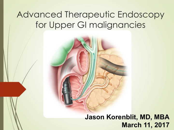
SLIDE 1
Advanced Therapeutic Endoscopy for Upper GI malignancies
Jason Korenblit, MD, MBA March 11, 2017
SLIDE 2
2
Disclosures
´ Financial disclosures: None, unfortunately.
SLIDE 3
Educational Objectives ´ Available Devices and Modalities ´ Staging EUS ´ Therapeutic EUS ´ Endoscopic mucosal resection ´ ERCP and Cholangioscopy ´ Deep enteroscopy
SLIDE 4
Endoscopic Ultrasound
Radial scope – diagnostic only Curvilinear scope – diagnostic and therapeutic Probe-based – diagnostic only
SLIDE 5
EUS and staging
SLIDE 6 Esophageal Cancer Staging
´ EUS is the most accurate modality for locoregional T staging
´ 80-90% ´ In one metaanalysis, AUROC 0.96 ´ T1(a) lesions can be endoscopically resected
´ Nodal staging
´ Endoscopic criteria
´ width greater than 10 mm ´ round shape ´ smooth border ´ echo-poor pattern
´ 80-100% predictor of nodal mets when all four present.
SLIDE 7
SLIDE 8
Restaging After Neoadjuvant Therapy
Restaging after resection: Sen 92% Spec 96%
SLIDE 9
Thoracic EUS
´ Mediastinum
´ Lymphoma ´ Thymus cancer ´ Metastatic Disease
´ Lung tumors
´ Posterior and inferior mediastinum
´ EBUS better for superior and anterior
´ Hilar lesions near the esophagus
´ Sampling of pleural effusions ´ Node Sampling ´ Celiac staging
SLIDE 10
EUS in the Stomach
´ T1 vs T2
´ Sen 85%, Spec 90% ´ T1 tumor limited to submucosa or shallower amenable to endoscopic resection
´ T1/2 vs T3/4 tumor
´ Sen 86, Spec 90%
´ EUS imaging alone can detect nodes as well as CT but DOES NOT predict malignancy well
´ Specificity only 67%, heterogeneous studies ´ EUS/FNA of nodes preferred
´ Stromal lesions benefit from EUS to determine what layers they arise in
´ EUS/FNA for tissue
SLIDE 11
Hypoechoic lesion arising in the muscularis propia Needle in mass
SLIDE 12 Endoscopic Mucosal Resection
´ Esophagus
´ Barrett’s nodule ´ T1A adenocarcinoma
´ Less than 2cm ´ Less than 1/3 circumference of the lumen
´ Stomach
´ Stromal tumors and adenocarcinoma limited to submucosa
´ 3% recurrence in Japanese literature
´ Large lesions may benefit from Endoscopic submucosal dissection for en bloc resection
´ Duodenum
´ Much higher perforation rate
´ Colon
´ Very amenable but watch for signs of muscularis propia invasion
´ Failure to lift ´ Depression
SLIDE 13
Traditional Saline/Fluid Lift
SLIDE 14
Cap-assisted Saline Lift
SLIDE 15
Band-assisted EMR
SLIDE 16
Defect Closure
Clips Endoscopic Suturing
SLIDE 17
Pancreatic EUS
´ Staging data is very mixed in pancreas
´ T staging accuracy 63-67% ´ N staging accuracy 44-66%
´ Assessing vascular invasion
´ Data actually shows EUS getting WORSE over time
´ Likely due to quality of the studies
´ Much better for portal vein and splenic art/vein than SMA/SMV
´ EUS is better than CT for detection of small lesions (98% vs 86%), about the same for nodes (about 47%), about the same for determining resectability (accuracy around 90%)
SLIDE 18 Who needs an FNA?
´ Tissue prior to Chemo/XRT
´ Advanced disease/Nodes ´ Poor surgical candidate ´ Metastatic disease
´ Resectable patient with small tumor (and no jaundice?)
´ Traditionally right to surgery ´ FNA can be helpful
´ Neuroendocrine tumors ´ Lymphoma ´ Autoimmune pancreatitis ´ Chronic pancreatitis
´ Sens 92%, Spec 96% ´ Accuracy 91% in patients without risk for chronic pancreatitis
SLIDE 19
Normal HPB Anatomy
SLIDE 20
Traditional ERCP vs Cholangioscopy
´ Fluoroscopy only ´ Fluoroscopic guidance/blind sampling ´ Wire manipulation using 2D image only ´ Needs Contrast ´ Video Cholangioscopy adds ´ Direct biopsy/sampling ´ Direction of wire under combined visual and fluoroscopic guidance ´ Can limit contrast injection and possibly prevent infection ´ Ability to discern malignancy vs benign disease using imaging ´ Precise guidance of certain therapies
SLIDE 21
Pancreaticobiliary Cancers
´ Liver tumors
´ Primary HCC ´ Metastatic tumors
´ Cholangiocarcinomas
´ Hilar tumors ´ Extrahepatic ´ Intrahepatic
´ Pancreatic ´ Gallbladder ´ Ampullary
SLIDE 22 ERCP with Brush Cytology
AUTHOR / YEAR PATIENTS N SENSITIVITY (%) SPECIFICITY (%)
Ponchon, 1995 233 35% 97% Lee, 1995 149 37% 100% Ornellas, 2006 50 40% 100% Jailwala, 1999 133 30% 100% TOTAL 565 36% 99% Fukuda – Gastrointestinal Endoscopy Volume 62, No. 3, 2005 “The most convenient and widely used method for tissue sampling from the biliary stricture and a filling defect is brushing
- cytology. However, the sensitivity and the negative prediction
values are insufficient for deciding the treatment plan.”
SLIDE 23 ERCP with Fluoroscopically Guided Biopsy
AUTHOR / YEAR PATIENTS N SENSITIVITY (%) SPECIFICITY (%)
Ponchon, 1995 128 43% 97% Schoefl, 1997 103 65% 100% Jailwala, 2000 133 37% 100% TOTAL 364 48% 99% Ponchon - Gastrointestinal Endoscopy December; 42(6):565-72 “Conclusions: Endobiliary sampling is technically difficult and has a limited sensitivity for the diagnosis of malignant biliary stenosis.”
SLIDE 24
Intraductal Papillary Mucinous Tumor
´ Mucinous tumor formed in the cells lining pancreatic duct ´ Typically causes pancreatic duct to dilate which obstructs flow of pancreatic juices to duodenum ´ Precursor to cancer
IPMT
SLIDE 25
Catheter-Based Cholangioscopy
´ Single operator ´ Similar size to mother/daughter scope ´ Video imaging ´ 1.2mm accessory channel ´ Disposable ´ 4 way tip deflection
SLIDE 26
Clinical Application of Technology
´ Stones
´ Choledocholithiasis ´ Intraductal stones ´ MPD stones
SLIDE 27
´ Strictures
´ Benign ´ Malignant
Clinical Application of Technology
SLIDE 28
Clinical Application of Technology
IPMN Biliary Anastomosis
SLIDE 29
Efficacy
´ ERCP with brushing alone or with fluoroscopic biopsy
´ Sensitivity 30%-50%, specificity 95%-99%
´ ERCP with Cholangioscopy alone
´ Meta-analysis of 8 prospective studies ´ Sensitivity 90%, specificity 87%
´ ERCP with cholangioscopy- directed biopsy
´ Sensitivity 69%, specificity 98%
´ Pancreatic strictures
´ IPMN’s detected in 95% of cases, benign strictures in 80%, cancers in 63% ´ Best for IPMN
SLIDE 30
Intraductal cholangiocarcinoma
SLIDE 31
Potential Complications of Cholangioscopy
´ Pancreatitis ´ Perforation ´ Hemorrhage ´ Hematoma ´ Septicemia/infection ´ Cholangitis ´ Allergic reaction to contrast medium
SLIDE 32
Small Bowel Evaluation
´ Video Capsule
´ Non-invasive ´ 60% detection rate in all lesions/bleeding sources ´ No therapy possible
´ Push Enteroscopy
´ Up to 80cm past the Ligament of Trietz ´ Colonoscope or dedicated enteroscope ´ Potentially therapeutic
SLIDE 33
Deep Enteroscopy
Balloon-Assisted Enteroscopy Spiral Overtube Enteroscopy
SLIDE 34
SLIDE 35
Deep Enteroscopy
´ Requires general anesthesia (usually) ´ Time consuming – 60 to 120 minutes ´ Up to 360cm past Ligament of Treitz antegrade ´ Up to 180cm from the ileocecal valve retrograde
´ 30% failure rate!
´ Possible to see the entire GI tract with both exams
´ 70-86% in Japan, 4-63% in US/Europe
´ Due to combination of experience and patient size
SLIDE 36
Summary and Recommendations
´ When in doubt, refer for EUS
´ Nodules ´ Extrinsic GI lesions
´ If it is within 7cm of the upper GI tract, we can hit it ´ Combination of EUS and EMR is effective for the management of early mucosal lesions and obviates surgery ´ Therapeutic ERCP can be used to establish diagnosis and palliate the patient ´ Deep enteroscopy can be used to obtain tissue reliably to the mid-small bowel
SLIDE 37 References
´ Osanai M, Itoi T, Igarashi Y, et al. Peroral video cholangioscopy to evaluate indeterminate bile duct lesions and preoperative mucosal cancerous extension: a prospective multicenter study. Endoscopy 2013; 45:635. ´ Lee YN, Moon JH, Choi HJ, et al. Direct peroral cholangioscopy using an ultraslim upper endoscope for management of residual stones after mechanical lithotripsy for retained common bile duct stones. Endoscopy 2012; 44:819. ´ Sasahira N, Isayama H, Nagano R, et al. Noncalcified pancreatic stone treated with electrohydraulic lithotripsy using SpyGlass pancreatoscopy. Endoscopy 2011; 43 Suppl 2 UCTN:E272. ´ Shah RJ, Chen YK. Transpapillary and percutaneous choledochoscopy in the evaluation and management of biliary strictures and stones. Techniques in Gastrointest Endosc 2007; 9:161. ´ Chen YK, Pleskow DK. SpyGlass single-operator peroral cholangiopancreatoscopy system for the diagnosis and therapy of bile-duct disorders: a clinical feasibility study (with video). Gastrointest Endosc 2007; 65:832. ´ Brauer BC, Chen YK, Shah RJ. Single-step direct cholangioscopy by freehand intubation using standard endoscopes for diagnosis and therapy of biliary diseases. Am J Gastroenterol 2012; 107:1030.