
SLIDE 1
- A. Holzinger
LV 709.049
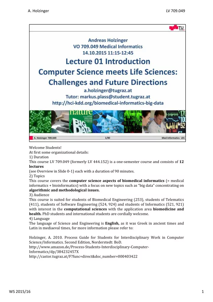
Welcome Students! At first some organizational details: 1) Duration This course LV 709.049 (formerly LV 444.152) is a one‐semester course and consists of 12 lectures (see Overview in Slide 0‐1) each with a duration of 90 minutes. 2) Topics This course covers the computer science aspects of biomedical informatics (= medical informatics + bioinformatics) with a focus on new topics such as “big data” concentrating on algorithmic and methodological issues. 3) Audience This course is suited for students of Biomedical Engineering (253), students of Telematics (411), students of Software Engineering (524, 924) and students of Informatics (521, 921) with interest in the computational sciences with the application area biomedicine and
- health. PhD students and international students are cordially welcome.