
4/18/2018 1
Pulmonary Embolism: Assessment, risk-stratification, and treatment plan for the
- utpatient management of
low-risk patients
Presentation by Joshua T. Wood, PharmD/PGY-1 Resident Providence St. Patrick Hospital; Missoula, MT Co-Investigators Jayme Hartzell, PharmD, MS, BCPS Disclosure Statement Financial
The investigators of this study have no financial conflicts of interest to disclose Non-Financial The primary investigator of this study has been provided published resources (including published studies, clinical pocket cards, and free trial vouchers) from Johnson and Johnson to aid in the development of this study. The primary investigator of this study has been provided published resources in the form of free trial vouchers from Bristol-Meyers Squibb
IRB Approval: Exempt status received January 11, 2018 Study sponsorship: None
Objectives
1) Compare and contrast the various validated tools for the identification of patients with pulmonary embolism 2) Distinguish between the different risk and mortality algorithms that exist and the merits of using multiple criteria for stratification
Background
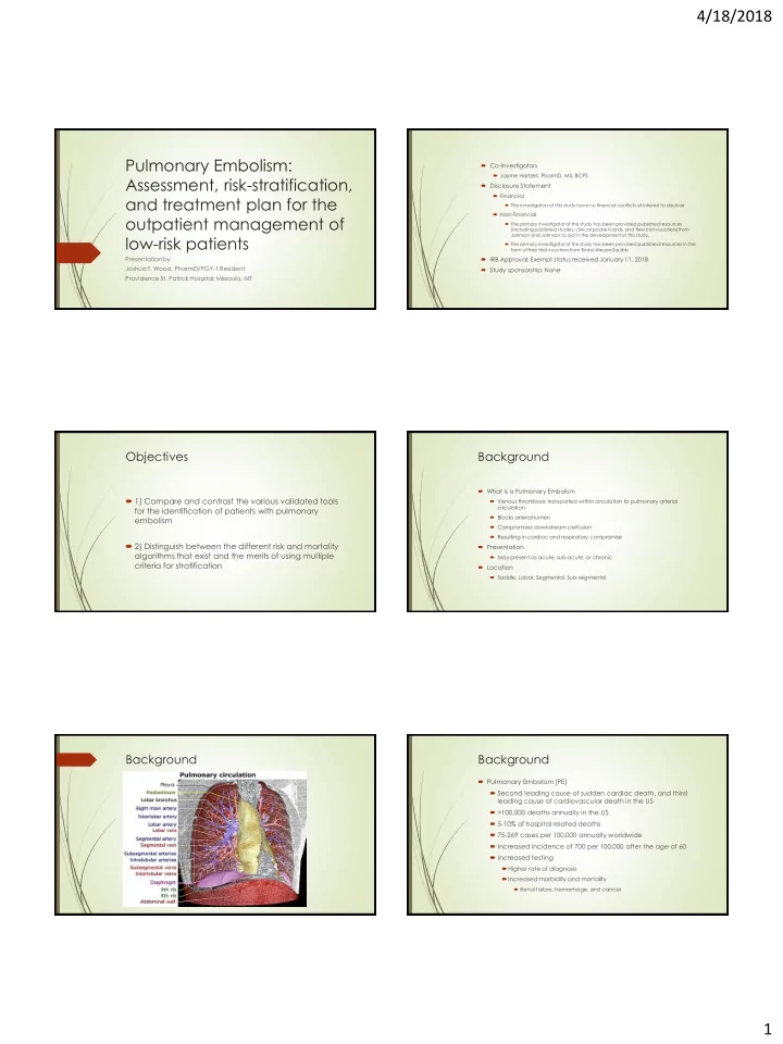
What is a Pulmonary Embolism Venous thrombosis transported within circulation to pulmonary arterial circulation Blocks arterial lumen Compromises downstream perfusion Resulting in cardiac and respiratory compromise Presentation May present as acute, sub-acute, or chronic Location Saddle, Lobar, Segmental, Sub-segmental
Background Background
Pulmonary Embolism (PE) Second leading cause of sudden cardiac death, and third leading cause of cardiovascular death in the US >100,000 deaths annually in the US 5-10% of hospital related deaths 75-269 cases per 100,000 annually worldwide Increased incidence of 700 per 100,000 after the age of 60 Increased testing Higher rate of diagnosis Increased morbidity and mortality
Renal failure, hemorrhage, and cancer