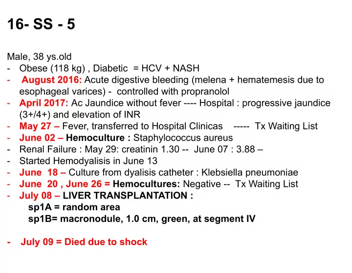
SLIDE 1 16- SS - 5
Male, 38 ys.old
- Obese (118 kg) , Diabetic = HCV + NASH
- August 2016: Acute digestive bleeding (melena + hematemesis due to
esophageal varices) - controlled with propranolol
- April 2017: Ac Jaundice without fever ---- Hospital : progressive jaundice
(3+/4+) and elevation of INR
- May 27 – Fever, transferred to Hospital Clinicas ----- Tx Waiting List
- June 02 – Hemoculture : Staphylococcus aureus
- Renal Failure : May 29: creatinin 1.30 -- June 07 : 3.88 –
- Started Hemodyalisis in June 13
- June 18 – Culture from dyalisis catheter : Klebsiella pneumoniae
- June 20 , June 26 = Hemocultures: Negative -- Tx Waiting List
- July 08 – LIVER TRANSPLANTATION :
sp1A = random area sp1B= macronodule, 1.0 cm, green, at segment IV
- July 09 = Died due to shock
SLIDE 2
SLIDE 3
Male, 38 ys.old. Obese , Diabetic,HCV + NASH = EXPLANT
SLIDE 4
Male, 38 ys.old. Obese , Diabetic,HCV + NASH = EXPLANT
SLIDE 5
Male, 38 ys.old. Obese , Diabetic,HCV + NASH = EXPLANT
SLIDE 6
Male, 38 ys.old. Obese , Diabetic,HCV + NASH = EXPLANT
SLIDE 7
Male, 38 ys.old. Obese , Diabetic,HCV + NASH = EXPLANT
SLIDE 8
Male, 38 ys.old. Obese , Diabetic,HCV + NASH = EXPLANT
SLIDE 9
Male, 38 ys.old. Obese , Diabetic,HCV + NASH = EXPLANT
SLIDE 10
Male, 38 ys.old. Obese , Diabetic,HCV + NASH = EXPLANT
SLIDE 11
Male, 38 ys.old. Obese , Diabetic,HCV + NASH = EXPLANT K7
SLIDE 12
Male, 38 ys.old. Obese , Diabetic,HCV + NASH = EXPLANT K 19
SLIDE 13
Male, 38 ys.old. Obese , Diabetic,HCV + NASH = EXPLANT
CD34
SLIDE 14
Male, 38 ys.old. Obese , Diabetic,HCV + NASH = EXPLANT Glutamine-synthase
SLIDE 15
Male, 38 ys.old. Obese , Diabetic,HCV + NASH = EXPLANT Arginin-Succinate-Synthase -ASS-1
SLIDE 16
Male, 38 ys.old. Obese , Diabetic,HCV + NASH = EXPLANT
ASS-1 GLUT-SYNT
SLIDE 17
MACRONODULE AT EXPLANT
SLIDE 18
MACRONODULE AT EXPLANT
SLIDE 19
MACRONODULE AT EXPLANT
SLIDE 20
MACRONODULE AT EXPLANT ASS-1 Glut-Synth
SLIDE 21
Male, 38 ys.old. Obese , Diabetic,HCV + NASH = EXPLANT
CONCLUSION
Advanced cirrhosis (4C)(Hepatitis C pattern prevailed) High septal angiogenesis and parenchymal vessel dilatation, “moderate chronic hepatitic type activity” High ACLF-Type activity” (High ductular reaction; High ductular cholestasis and inflammation ; confluent hepatic necrosis) (Type 1- severe – Rastogi et al, 2011) ACLF-related lesions especially intense in the Macro-Regenerative Nodule measuring 1.0 cm in segment IV.
SLIDE 22 16- SS - 5
Male, 38 ys.old
- Obese (118 kg) , Diabetic = HCV + NASH
- August 2016: Acute digestive bleeding (melena + hematemesis due to
esophageal varices) - controlled with propranolol
- April 2017: Ac Jaundice without fever ---- Hospital : progressive jaundice
(3+/4+) and elevation of INR
- May 27 – Fever, transferred to Hospital Clinicas ----- Tx Waiting List
- June 02 – Hemoculture : Staphylococcus aureus
- Renal Failure : May 29: creatinin 1.30 -- June 07 : 3.88 –
- Started Hemodyalisis in June 13
- June 18 – Culture from dyalisis catheter : Klebsiella pneumoniae
- June 20 , June 26 = Hemocultures: Negative -- Tx Waiting List
- July 08 – LIVER TRANSPLANTATION :
sp1A = random area sp1B= macronodule, 1.0 cm, green, at segment IV
- July 09 = Died due to shock
SLIDE 23
Rastogi A... Liver histology as predictor of outcome in patients with acute-on-chronic liver failure (ACLF) Virchows Arch (2011) 459:121–127
Univariate analysis of correlation of liver histology with outcome in patients of ACLF
SLIDE 24 Lefkowitch J - CHOLANGITIS LENTA in SEPSIS
Scheuer´s Liver Biopsy Interpretation, 2010,pg 57: "Septicaemia uncommonly is associated with a particular form of histological cholangitis principally affecting the canals of Hering . Affected ductules are dilated and filled with inspissated bile. Neutrophils acccumulate around and sometimes within
- them. Larger ducts may be affected, as may the
periportal parenchyma in which bile is seen in dilated bile canaliculi. Theses changes are easily confused with those of large bile-duct obstruction, but in obstruction the inspissated bile in the canals of Hering is not a feature, unless there is concomitant
- sepsis. Sepsis more often gives rise to widesperad
canalicular cholestasis...
SLIDE 25 Lefkowitch JH. Bile ductular cholestasis: an ominous histopathologic sign related to sepsis and "cholangitis lenta".Hum Pathol.1982;13:19-24.
An unusual form of intrahepatic cholestasis manifested by inspissated bile within dilated and proliferated portal and periportal bile ductules was seen in liver biopsy and autopsy specimens from three
- patients. Features of sepsis and severe systemic illness with
jaundice dominated their clinical presentations, and no autopsy evidence of large bile duct obstruction could be found. This lesion may be related to the old entity, "cholangitis lenta," a form of chronic sepsis associated with biliary tract inflammation in the absence of demonstrable extrinsic obstruction. Identification of this pattern of cholestasis in liver biopsy specimens is useful in certain patients who may be a great risk of mortality and who require serious clinical attention directed toward elucidating a source for sepsis as well as aggressive management of
SLIDE 26
Male, 38 ys.old. Obese , Diabetic,HCV + NASH = EXPLANT
SLIDE 27
INTRA-HEPATIC BILIARY SYSTEM INTRA-HEPATIC BILIARY SYSTEM
Sinusoid Microvilli
Hepatocyte
Microsome
Biliary Canaliculi
interlobular Duct (15-100 u) Septal Duct (>100 u) Canal of Hering Ductule/Cholangiole Portal Tract
LIM 14-LIVER PATHOLOGY HC-FMUSP, 2018 LIM 14-LIVER PATHOLOGY HC-FMUSP, 2018
Biliary Canaliculi Canals of Hering (CoH) Biliary Ductules (Cholangioles) Biliary Ducts : Interlobular: Small: 15-40m Intermediate: 40-100m Septal: > 100m Large biliary ducts: 3rd generation : 300-400m 2nd generation : 400-800m 1st generation : > 800m (hepatic right and left)
SLIDE 28
Male, 38 ys.old. Obese , Diabetic,HCV + NASH = EXPLANT
SLIDE 29
Male, 38 ys.old. Obese , Diabetic,HCV + NASH = EXPLANT
SLIDE 30
Male, 38 ys.old. Obese , Diabetic,HCV + NASH = EXPLANT
SLIDE 31 Jindal A,Rastogi A, Sarin S:
- Exp. Rev. Gastroenterol & Hepatology, 2016
If acute injury is controlled, recovery is more likely in ACLF In the first 1–2 weeks after injury onset,the patient can develop sepsis. This intervening period is a ‘golden window’ for treatment
SLIDE 32
Acute-on–chronic liver failure 2018: a need for (urgent) liver biopsy?
Expert Review of Gastroenterology & Hepatology 2018
The International Liver Pathology Study Group
Dirk J. van Leeuwen, Venancio Alves,Charles Balabaud Prithi S. Bhathal, Paulette Bioulac-Sage, Romano Colombari, James M. Crawford, Amar Dhillon, Linda Ferrell, Ryan Gill, Maria Guido, Prodromos Hytiroglou, Yasuni Nakanuma, Valerie Paradis, Pierre Emmanuel Rautou, Christine Sempoux, Dale C. Snover, Neil D. Theise, Swan N. Thung, Wilson M.S. Tsui & Alberto Quaglia
SLIDE 33
The International Liver Pathology Study Group – Expert Review of Gastroenterology & Hepatology 2018
SLIDE 34 Pathology
- Prof. Venâncio A. F. Alves
- Dr. Evandro S. Mello
Dra Fabiana Lima Dr Ryan Tanigawa
- Dr. Renan Ribeiro Dr. Sebastião Martins
- Dr. Dafne Andrade Dr.Arthur Volpatto
Hepatology
- Prof. Flair J. Carrilho
- Prof. Suzane K. Ono-Nita Prof. Alberto Q. Farias
- Prof.Claudia PM. Oliveira Prof. Eduardo Cancado
- Dr. Denise C. Paranaguá Vezozzo
- Dr Mario Guimarães Pessoa Dra. Aline L. Chagas
- Dra. Regiane S. M. Alencar
- Dra. Karla Toda
Hospital das Clínicas Faculdade de Medicina da Universidade de São Paulo
SLIDE 35 After mitigating the acute injury, spontaneous recovery is more likely in those with ACLF than ESLD due to a higher baseline hepatic reserve. After acute insult or injury, the condition of a patient with ACLF is likely to rapidly deteriorate . In the first 1–2 weeks after injury onset,the patient could also develop sepsis. This intervening period is a therapeutic ‘golden window’ to ameliorate the acute injury, supplement liver regeneration, to modulate the patient’s immune response to prevent sepsis and to prevent multiorgan failure and death.
Jindal A,Rastogi A, Sarin S:
- Exp. Rev. Gastroenterol & Hepatology, 2016