10/28/2011 Reafference Principle Holst E. von and Mittelstaedt H. ( - - PDF document

10 28 2011
SMART_READER_LITE
LIVE PREVIEW

10/28/2011 Reafference Principle Holst E. von and Mittelstaedt H. ( - - PDF document

10/28/2011 Reafference Principle Holst E. von and Mittelstaedt H. ( 1950 ) Da;. Reafferenzprincip. L36. EXPECTATION GENERATORS Naturwissenschaften 37, 464 476. October 28, 2011 C. D. Hopkins Much of sensory processing involves the


slide-1
SLIDE 1

10/28/2011 1

  • L36. EXPECTATION GENERATORS

October 28, 2011

  • C. D. Hopkins

“Much of sensory processing involves the generation of expectations or predictions about sensory input, and subsequent removal of such expectations from the sensory inflow.” Bell, C (1997) Brain, Behavior, Evolution 50 (suppl.) 17-31.

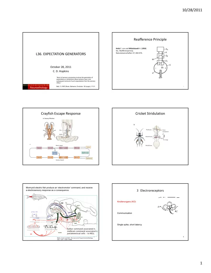
Holst E. von and Mittelstaedt H. (1950) Da;. Reafferenzprincip. Naturwissenschaften 37, 464‐476.

Reafference Principle

2

Crayfish Escape Response Cricket Stridulation

Mormyrid electric fish produce an ‘electromotor’ command, and receive a electrosensory response as a consequence.

5 Meek, Grant and Bell. The Journal of Experimental Biology 202, 1291–1300 (1999)

bulbar command associated n. midbrain command associated n. juxtalemniscal cells – to NELL

3 Electroreceptors

Knollenorgans (KO)

6

Communication Single spike, short latency

slide-2
SLIDE 2

10/28/2011 2

  • C. C. Bell and K. Grant

7

8

XU-FRIEDMAN, M. A. & HOPKINS, C. D., 1999.- J. Exp. Biol., 202:1311-1318. FRIEDMAN, M.A. & HOPKINS, C.D. 1998 – J. Neurosci.18:1171-1185.

9

XU-FRIEDMAN, M. A. & HOPKINS, C. D., 1999.- J. Exp. Biol., 202:1311-1318. FRIEDMAN, M.A. & HOPKINS, C.D. 1998 – J. Neurosci.18:1171-1185.

Knollenorgans blank inputs by inhibition

  • -Primary afferent (blue)

terminates on nELL cell (yellow) with

  • -large calyx-like

synapses (electrotonic).

  • -fibers from EOD

EOD command ipsp in nELL cell afferent 10

fibers from EOD command (eocd) produce spikes that arrive at same time that EOD would be fired.

  • -sharp inhibition

IPSP blocks spike at the time when EOD is expected

  • - removes expected

EOD

EOD

command

Mormyromasts (morm) Active electrolocation.

2 Mormyromasts

11

Burst duration code Ampullary (ao) Respond to D.C. stimuli

  • 3. Ampullary Receptors (=D.C. receptors)

12

Passive electrolocation of prey. Frequency code.

slide-3
SLIDE 3

10/28/2011 3

A Modifiable Efference Copy

13

Recording setup: 1) mormyrid electric fish 2) curarize electric fish – silence the electric organ 3) record command signal in tail 4) stimulate artificially, using command 5) record in ampullary electrosensory area of ELL

Bell, CC (1982)

14 Meek, Grant and Bell. The Journal of Experimental Biology 202, 1291–1300 (1999)

Principal Cell Plasticity

EOD command

15

Principal Cell Plasticity

EOD command

16

Modifiable Negative Image in histogram form

17

The negative image works for any stimulus delay after the command

18

COMMAND COMMAND

slide-4
SLIDE 4

10/28/2011 4

The negative image compensates stimulus applied anywhere on body surface

19

All recording from same ELL cell; stimulus applied to A, B, C, or D

Electrosensory Plasticity in Gymnotiform Fishes

20

Apteronotus leptorhynchus Eigenmannia virescens

Bastian: Sensory Consequence of Movement of Tail

Experimental paradigm: 1) Tail movement controlled by motor. 2) Electric discharge continues d ti l t

21

and stimulates electroreceptors. 3) Electrode near skin monitors the EOD amplitude

Propioceptors Monitor Tail Position

22

Electrophysiological recordings from propioceptors in the EGp of Apteronotus in response to tail bending experiment. Propioceptors monitor all positions of tail bend. (increases, decreases, middle).

Electrosensory Feedback From Tail Bending is Cancelled in ELL

23

The cancellation is modifiable over time.

If tail is not bent during artificial Amplitude modulation, no effect

24

slide-5
SLIDE 5

10/28/2011 5

Effect of Global EOD on Local Electrosensory Response

A) Response of basilar pyramidal cell to whole body AM.

25

y B) Body AM plus local AM B4) Body AM alone, local AM off.

Now, with removal of EOD

x Now, the EOD is silenced to prevent global stimulation.

26

A) Tail is moved back and forth to provide the proprioceptive cue. B) Local AM causes stimulation of electroreceptors. C) After pairing, response to tail movement alone, no AM

27

The Basic Architecture

1. Bell, C. C., Han, V. and Sawtell, N. B. (2008). Cerebellum-like structures and their implications for cerebellar function. Annu R N i 31 1 24

Methods (continued)

Identification of Mossy Fibers in Egp. 1) Previous studies by Bell (1991) recorded from cells in EGp with similar properties and found * no synaptic activity, *spikes arising directly from baseline. 2) Several fills with biocytin (3 from proprioceptive input, 2 with EODc input) 3) No neurons responded to both EODc and propioception. 4) Pairing of EODCD synaptic activity and narrow spike activity by averaging responses before and after EODCD, narrow spikes removed.

Anatomy

Injection of biotinylated dextran into molecular layer of ELL labels: MG cells (gabaergic cells in ELL) granule cells which project via parallel fibers to MGcell dendrites.

slide-6
SLIDE 6

10/28/2011 6

Many Mossy Fibers provide propioceptive sensory information from tail.

(most common type of unit)

responds to tail bend, not EODc. Spike rate unchanged by EOD. Firing rate is 20-150 Hz. Has a best tail angle

most preferred

  • extremes. A few

intermediate l

g Spike rate encodes tail angle shown for 3 units that prefer contralateral bend, 3 that prefer ipsilateral bend. Some proprioceptor units respond to schnauzenorgan displacement, trunk displacement or fin.

angles

Some mossy fibers respond to EOD command

Putative MossyFiber cell responding to EODc. No spontaneous activity. Response precedes EOD.

  • ccasion

3 units showing differing latencies of responses. Stereotyped rasters. Summed histograms of spikes per command from 30 such units. Active for up to 400 msec after EODc

  • ccasion

al cell had burst, then long latency burst.

33

Circuitry in the ELL

34

Theory to Explain Negative Image

Sensory input arrives from below and stimulates pyramidal cell to fire. Predictive input comes from above through parallel fibers.

35

Synaptic modification changes strength of predictive input as a consequence of simultaneous action.

Recording Plasticity in Slice Preparation

Recording intracellularly from an ELL cell from mormyromast region

  • f mormyrid ELL.

36

Stimulate parallel fibers with two sets of electrodes in molecular layer of ELL.

slide-7
SLIDE 7

10/28/2011 7

Spike timing dependent plasticity is anti-hebbian. If the broad spike occurs after the EPSP, the synaptic weight is decreased. If the broad spike occurs before the EPSP, it strengthens. This sculpts a negative image of the expected input.

Percentage change in excitatory postsynaptic potential (EPSP) amplitude plotted against the delay between EPSP onset and the broad spike peak during pairing. A negative delay with regard to the previous spike and a positive delay with regard to the following spike were present for each pairing. The shorter of these two delays is

  • plotted. Filled circles, significant changes; open circles, non-significant changes (at P<0.01).

animation rule

Other Examples of Removing Expected Inputs

  • Adaptation of receptors or neurons to maintained

stimuli removes responses to constant stimulus. L t l i hibiti t d l l

38

  • Lateral inhibition: remove expected mean levels
  • ver space.”

Both methods use simple non-plastic cellular changes (self inhibition, intra-cellular inhibition).