1 UCSF Vascular & Endovascular Symposium 2015 UCSF - - PowerPoint PPT Presentation

1
SMART_READER_LITE
LIVE PREVIEW

1 UCSF Vascular & Endovascular Symposium 2015 UCSF - - PowerPoint PPT Presentation

UCSF Vascular & Endovascular Symposium 2015 UCSF Vascular & Endovascular Symposium 2015 UC SF UC SF TBAD Anatomy & Pathophysiology TBAD Anatomy & Pathophysiology DISCLOSURES INDIVIDUAL CURRENT UNDERSTANDING:


slide-1
SLIDE 1

1

UCSF Vascular & Endovascular Symposium 2015

TBAD Anatomy & Pathophysiology

UCSF

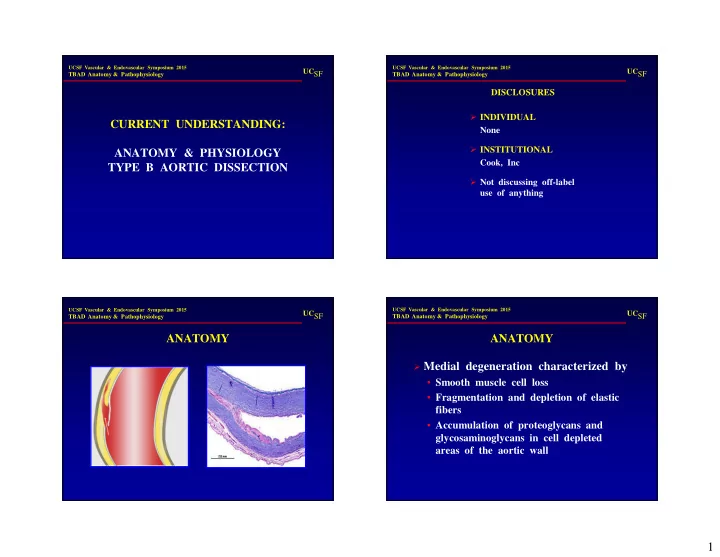
CURRENT UNDERSTANDING: ANATOMY & PHYSIOLOGY TYPE B AORTIC DISSECTION

UCSF Vascular & Endovascular Symposium 2015

TBAD Anatomy & Pathophysiology

UCSF

DISCLOSURES INDIVIDUAL None INSTITUTIONAL Cook, Inc Not discussing off-label use of anything

UCSF Vascular & Endovascular Symposium 2015

TBAD Anatomy & Pathophysiology

UCSF

ANATOMY

UCSF Vascular & Endovascular Symposium 2015

TBAD Anatomy & Pathophysiology

UCSF

ANATOMY

Medial degeneration characterized by

  • Smooth muscle cell loss
  • Fragmentation and depletion of elastic

fibers

  • Accumulation of proteoglycans and

glycosaminoglycans in cell depleted areas of the aortic wall

slide-2
SLIDE 2

2

UCSF Vascular & Endovascular Symposium 2015

TBAD Anatomy & Pathophysiology

UCSF

ANATOMY

Progression results from interplay of

  • Hemodynamic stress
  • Aortic injury
  • Chronic inflammation
  • Genetic propensity
  • Epidemiologic risk factors

UCSF Vascular & Endovascular Symposium 2015

TBAD Anatomy & Pathophysiology

UCSF

ANATOMY

Media consists of elastic fibers and vascular

smooth muscle cells, interconnected by collagen fibers, proteoglycans, glycosaminoglycans and various adhesive proteins

When functioning normally, these ECM

components

  • Impart elasticity & tensile strength
  • Sequester growth factors
  • Form structural interactions

UCSF Vascular & Endovascular Symposium 2015

TBAD Anatomy & Pathophysiology

UCSF

ANATOMY

All aortic media components show defects

  • Elastin—fibers, microfibrils, associated

proteins & interface proteins

  • Collagen—increased expression, disordered

deposition

  • Smooth muscle cells

Apoptosis Phenotype change

  • PG / GAG—content, function

UCSF Vascular & Endovascular Symposium 2015

TBAD Anatomy & Pathophysiology

UCSF

ANATOMY

All the usual inflammatory markers are

present (T-cells, macrophages, cytokines)

Hypoxia and increased oxidative stress An excess of proteases (including MMPs) Pathologic signalling

  • TGFβ
  • Angiotensin II
slide-3
SLIDE 3

3

UCSF Vascular & Endovascular Symposium 2015

TBAD Anatomy & Pathophysiology

UCSF

Percentage 60% 10-15% 25-30% Type DeBakey I DeBakey II DeBakey III Stanford A Stanford B Proximal Distal

ANATOMY

UCSF Vascular & Endovascular Symposium 2015

TBAD Anatomy & Pathophysiology

UCSF

ANATOMY

Golledge et al, Lancet, 2008 Svensson et al, Circulation, 1999

UCSF Vascular & Endovascular Symposium 2015

TBAD Anatomy & Pathophysiology

UCSF

ANATOMY

UCSF Vascular & Endovascular Symposium 2015

TBAD Anatomy & Pathophysiology

UCSF

ANATOMY

INTIMAL TEAR

Location

  • Outer curve

less likelihood of retrograde extension possibly less likelihood of late enlargement

  • Inner curve

greater likelihood of retrograde extension (only 15% with NO arch or ascending aortic involvement)

slide-4
SLIDE 4

4

UCSF Vascular & Endovascular Symposium 2015

TBAD Anatomy & Pathophysiology

UCSF

ANATOMY

INTIMAL TEAR

Size / Number

  • Less likelihood of late enlargement

small entry tear (<10 mm) multiple tears

  • More likelihood of late enlargement

large entry tear single / few tears no distal tear

UCSF Vascular & Endovascular Symposium 2015

TBAD Anatomy & Pathophysiology

UCSF

ANATOMY

INTIMAL TEAR Caution

How imaged How measured Modeling vs in vivo

UCSF Vascular & Endovascular Symposium 2015

TBAD Anatomy & Pathophysiology

UCSF

ANATOMY

AORTIC DIAMETER

More likelihood of late enlargement

  • Overall initial aortic diameter ≥ 40 mm
  • Proximal descending thoracic aortic false

lumen ≥ 22 mm

  • Initial diameter of the mid-thoracic aorta

UCSF Vascular & Endovascular Symposium 2015

TBAD Anatomy & Pathophysiology

UCSF

ANATOMY LUMEN LOCATION, SHAPE & FLOW

Increased Risk True lumen

Elliptical shape

False lumen

Patent Partially thrombosed Saccular Inner curve

Decreased Risk True lumen

Circular lumen

False lumen

Thrombosed Outer curve

slide-5
SLIDE 5

5

UCSF Vascular & Endovascular Symposium 2015

TBAD Anatomy & Pathophysiology

UCSF

PHYSIOLOGY

HEMODYNAMICS

Importance of false lumen pressure

  • BP control

Onitsuka et al, J Thor Surg, 2003

UCSF Vascular & Endovascular Symposium 2015

TBAD Anatomy & Pathophysiology

UCSF

PHYSIOLOGY

HEMODYNAMICS

Importance of false lumen pressure

  • HR control

Kodama et al, Circulation, 2008

UCSF Vascular & Endovascular Symposium 2015

TBAD Anatomy & Pathophysiology

UCSF

PHYSIOLOGY

HEMODYNAMICS

Static Obstruction Dynamic Obstruction

UCSF Vascular & Endovascular Symposium 2015

TBAD Anatomy & Pathophysiology

UCSF

PHYSIOLOGY

HEMODYNAMICS

Static Obstruction

  • aortic stent-graft
  • branch stent
  • thrombectomy

Dynamic Obstruction

  • aortic stent-graft
  • fenestration
slide-6
SLIDE 6

6

UCSF Vascular & Endovascular Symposium 2015

TBAD Anatomy & Pathophysiology

UCSF

PATHOPHYSIOLOGY

GENETICS

50 year old man, with Type B dissection, mild (one- drug) antecedent hypertension, no abnormal morphometrics, a cousin who had an aortic aneurysm (not dissection). He probably has

  • A. Hypertension-related sporadic TAD
  • B. Some form of Marfan’s
  • C. Some other genetic abnormality
  • D. Bad luck

H y p e r t e n s i

  • n
  • r

e l a t e d s p . . . S

  • m

e f

  • r

m

  • f

M a r f a n ’ s S

  • m

e

  • t

h e r g e n e t i c a b . . . B a d l u c k

51% 14% 31% 3%

UCSF Vascular & Endovascular Symposium 2015

TBAD Anatomy & Pathophysiology

UCSF

PATHOPHYSIOLOGY

GENETICS

Syndromic

DeBacker et al, Ann Cardiothor Surg, 2013

UCSF Vascular & Endovascular Symposium 2015

TBAD Anatomy & Pathophysiology

UCSF

PATHOPHYSIOLOGY

GENETICS

Syndromic

SYNDROME GENE LOCATION Marfans Fibrillin 1 Mostly ascending aorta, aortic root; few descending aorta Loeys-Dietz 1 TGFBR1/2 Aortic root Ehlers-Danlos IV COL3A1 Mostly thoracic aorta and abdominal aorta Aneurysm-Osteoarthritis SMAD3 Mostly ascending aorta Turners syndrome 46X 15% are Type B eNOS 4a/b polymorphism 90% Type A

UCSF Vascular & Endovascular Symposium 2015

TBAD Anatomy & Pathophysiology

UCSF

PATHOPHYSIOLOGY

GENETICS

Non-Syndromic

genes related to smooth muscle contractile function

Isolated / sporadic

DeBacker et al, Ann Cardiothor Surg, 2013

slide-7
SLIDE 7

7

UCSF Vascular & Endovascular Symposium 2015

TBAD Anatomy & Pathophysiology

UCSF

PATHOPHYSIOLOGY

GENETICS

Non-Syndromic

  • Estimated that 20% of TAAD have a

genetic component

  • Autosomal dominant
  • Interfamilial variability
  • Variable penetrance
  • Variable severity

UCSF Vascular & Endovascular Symposium 2015

TBAD Anatomy & Pathophysiology

UCSF

PATHOPHYSIOLOGY

GENETICS

ACTA2 (12-21%)

  • About 50% have aortic events

Thoracic dissection 25% mortality Type A > Type B Age of Type B < age at Type A

  • Arterial occlusive disease
  • Livedo reticularis
  • Dissect at diameter of 4.5-5.0 cm

TGFβ2 (4%)

UCSF Vascular & Endovascular Symposium 2015

TBAD Anatomy & Pathophysiology

UCSF

PATHOPHYSIOLOGY

GENETICS

MYH11 (1-2%)

  • Patent ductus
  • Dissect at diameter of 4.0-4.5 cm

MLYK dissect with no enlargement FTAAD5 causes proteoglycan deposition

UCSF Vascular & Endovascular Symposium 2015

TBAD Anatomy & Pathophysiology

UCSF

PATHOPHYSIOLOGY

GENETICS TGFβ Arteriopathies

Marfan’s abnormal fibrillin-1 failed sequestration of TGFβ complex, resulting in increased active TGFβ & TGFβ signalling Loeys-Dietz TGFBR1 & 2 increased active TGFβ Aneurysm/ SMAD gene abnormalities in the TGFβ2 ligand Osteoarthritis ACTA2 increased TGFβ signalling MYH11 increased TGFβ signalling FTAAD5 increased PG deposition, ? caused by TGFβ signalling

slide-8
SLIDE 8

8

UCSF Vascular & Endovascular Symposium 2015

TBAD Anatomy & Pathophysiology

UCSF

PATHOPHYSIOLOGY

GENETICS TGFβ Arteriopathies

Marfan’s abnormal fibrillin-1 failed sequestration of TGFβ complex, resulting in increased active TGFβ & TGFβ signalling Loeys-Dietz TGFBR1 & 2 increased active TGFβ Aneurysm/ SMAD gene abnormalities in the TGFβ2 ligand Osteoarthritis ACTA2 increased TGFβ signalling MYH11 increased TGFβ signalling FTAAD5 increased PG deposition, ? caused by TGFβ signalling

UCSF Vascular & Endovascular Symposium 2015

TBAD Anatomy & Pathophysiology

UCSF

PATHOPHYSIOLOGY

GENETICS

DeBacker et al, Ann Cardiothor Surg, 2013

UCSF Vascular & Endovascular Symposium 2015

TBAD Anatomy & Pathophysiology

UCSF

PATHOPHYSIOLOGY

GENETICS

Hiratzka et al, Circulation, 2010

UCSF Vascular & Endovascular Symposium 2015

TBAD Anatomy & Pathophysiology

UCSF

PATHOPHYSIOLOGY

GENETICS

Hiratzka et al, Circulation, 2010

slide-9
SLIDE 9

9

UCSF Vascular & Endovascular Symposium 2015

TBAD Anatomy & Pathophysiology

UCSF

PATHOPHYSIOLOGY

GENETICS

Hiratzka et al, Circulation, 2010

UCSF Vascular & Endovascular Symposium 2015

TBAD Anatomy & Pathophysiology

UCSF

PATHOPHYSIOLOGY

GENETICS

Hiratzka et al, Circulation, 2010

UCSF Vascular & Endovascular Symposium 2015

TBAD Anatomy & Pathophysiology

UCSF