
SLIDE 1
1
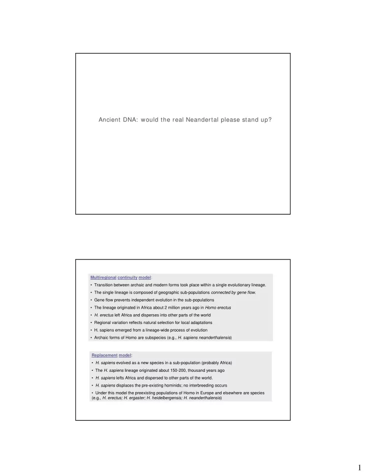
Ancient DNA: would the real Neandertal please stand up?
Multiregional continuity model:
- Transition between archaic and modern forms took place within a single evolutionary lineage.
- The single lineage is composed of geographic sub-populations connected by gene flow.
- Gene flow prevents independent evolution in the sub-populations
- The lineage originated in Africa about 2 million years ago in Homo erectus
- H. erectus left Africa and disperses into other parts of the world
- Regional variation reflects natural selection for local adaptations
- H. sapiens emerged from a lineage-wide process of evolution
- Archaic forms of Homo are subspecies (e.g., H. sapiens neanderthalensis)
Replacement model:
- H. sapiens evolved as a new species in a sub-population (probably Africa)
- The H. sapiens lineage originated about 150-200, thousand years ago
- H. sapiens lefts Africa and dispersed to other parts of the world.
- H. sapiens displaces the pre-existing hominids; no interbreeding occurs
- Under this model the preexisting populations of Homo in Europe and elsewhere are species