
SLIDE 1
1
1
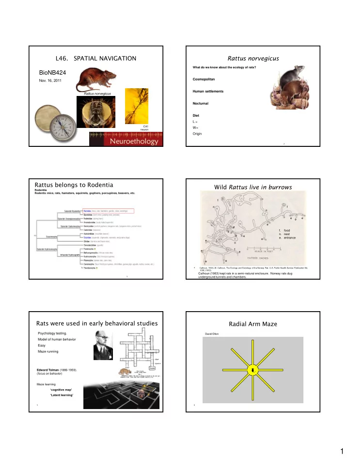
- L46. SPATIAL NAVIGATION
BioNB424
- Nov. 16, 2011
Rattus norvegicus
CA1 neuron
2
Rattus norvegicus
What do we know about the ecology of rats?
Cosmopolitan Human settlements Nocturnal Diet L = W= Origin
3
Rattus belongs to Rodentia
Rodentia Rodents: mice, rats, hamsters, squirrels, gophers, porcupines, beavers, etc.
Wild Rattus live in burrows
4
Calhoun (1963) kept rats in a semi-natural enclosure. Norway rats dug underground tunnels and chambers.
- f. food
- n. nest
- e. entrance
e e e e e e e e e e e e e e n n n f f f
Calhoun, 1963 J.B. Calhoun, The Ecology and Sociology of the Norway Rat. U.S. Public Health Service Publication No. 1008, (1963).
5
Rats were used in early behavioral studies
Psychology testing. Model of human behavior Easy Maze running
Edward Tolman (1886-1959). (focus on behavior) Maze learning ‘cognitive map’ ‘Latent learning’
6
Radial Arm Maze
David Olton