
SLIDE 1
10/16/2013 1
Atypical Wounds
Hollie Smith Mangrum, PT, DPT, CWS, FACCWS
Chronic Wounds
- Incidence of chronic wound in US is 6 million per year
- Majority ????
- 10% of lower extremity ulcers are due to less frequent
etiologies
- Inflammatory processes
- Infection
- Metabolic disorders
- Neoplasms
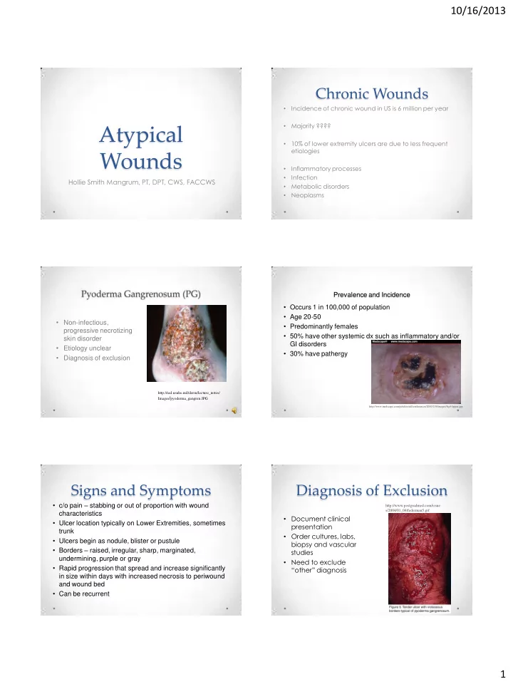
- Non-infectious,
progressive necrotizing skin disorder
- Etiology unclear
- Diagnosis of exclusion
http://rad.usuhs.mil/derm/lecture_notes/ Images/pyoderma_gangren.JPG
Prevalence and Incidence
- Occurs 1 in 100,000 of population
- Age 20-50
- Predominantly females
- 50% have other systemic dx such as inflammatory and/or
GI disorders
- 30% have pathergy
http://www.medscape.com/pi/editorial/conferences/2000/119/images/fig4-lipper.jpg
Signs and Symptoms
- c/o pain – stabbing or out of proportion with wound
characteristics
- Ulcer location typically on Lower Extremities, sometimes
trunk
- Ulcers begin as nodule, blister or pustule
- Borders – raised, irregular, sharp, marginated,
undermining, purple or gray
- Rapid progression that spread and increase significantly
in size within days with increased necrosis to periwound and wound bed
- Can be recurrent
Diagnosis of Exclusion
- Document clinical
presentation
- Order cultures, labs,
biopsy and vascular studies
- Need to exclude
“other” diagnosis
http://www.postgradmed.com/issue s/2004/01_04/federman5.gif