
SLIDE 1
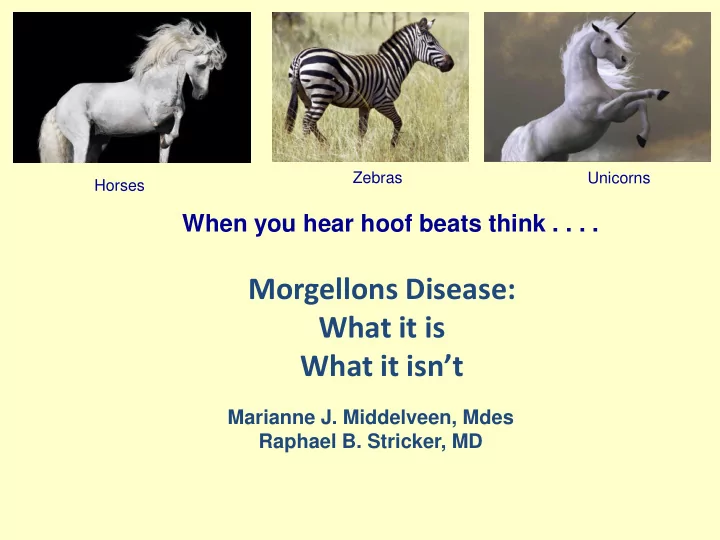
Morgellons Disease: What it is What it isn’t
Marianne J. Middelveen, Mdes Raphael B. Stricker, MD
When you hear hoof beats think . . . .
Horses Zebras Unicorns
What it is What it isnt Marianne J. Middelveen, Mdes Raphael B. - - PowerPoint PPT Presentation
Zebras Unicorns Horses When you hear hoof beats think . . . . Morgellons Disease: What it is What it isnt Marianne J. Middelveen, Mdes Raphael B. Stricker, MD Disclosure Statement Speaker: Marianne J. Middelveen, Mdes Consultant
Marianne J. Middelveen, Mdes Raphael B. Stricker, MD
Horses Zebras Unicorns
Consultant Microbiologist Atkins Veterinary Services
1682 – drawing by
“Inanimate, Levenloos”
Father of Microbiology
Sir Thomas Browne
Treponema pallidum spirochetal agent
1st Conference CEHMDF Cindy Casey Holman RN, Charles E Holman Matchbox and skin specimens Mary Leitao with her children
fakenews.com
Delusional disorder/Textile fibers. Peer-reviewed medical literature. Infectious etiology/ Biofibers of human origin. Peer-reviewed medical studies. Infectious etiology/ Filamentous organisms i.e. molds, insects, worms. Speculation, no evidence. Extraterrestrial origin/ Mystery fibers. Fantasy, no evidence. Bioengineering by nanites etc. / Man-made filaments. Fantasy, no evidence. Toxins/Filaments of silicone, dental adhesive, chemtrails, GMOs, mercury etc. Fantasy, no evidence.
ucmp.berkeley.edu mst3k.wikia.com
to the American Psychiatric Association’s DSM-5, used inappropriate case definitions, inadequate laboratory analysis, none of the studies used methods appropriate to find borreliosis.
Clinic, and 1 collaborative study by Centers for Disease Control and Prevention (CDC) and Kaiser Permanente (KP), all failed to perform lab analysis to detect spirochetes/borreliosis, all had flawed case definitions; fiber analysis was flawed and included analysis of contaminating cotton fibers.
based and that offer little or no original research evidence.
Image: healthymind.com
from skin and fluid (detected viable
independent laboratories provided corroborative evidence
microscopy (SEM) and transmission electron microscopy (TEM) were forwarded to the Electron Microscopy Facility, Department of Materials Science and Engineering, Clemson University, Anderson, South Carolina.
submitted samples – both in tissue and cultures.
SEM – skin culture
TEM – callus section
Spirochetes (dark brown/black) detected in Dieterle-stained histological sections of Morgellons tissue
standard silver nitrate stains used to detect spirochetes.
fixed and processed for silver nitrate staining at Interscope Laboratories, Canoga Park, CA (ISL) and McClain Clinical Laboratories, Smithtown, NY (MCL).
lesions have larger spirochetal load.
Dieterle-stained cultured spirochetes
Helical spirochete MD histological section – callus Helical spirochete MD vaginal culture Immunostain MD callus section.
Single spirochete Immunostain MD skin culture.
Photos: Divya Burugu, University of New Haven
Human psoriasis skin
Human skin with normal bacterial flora
Positive control Bb B-31 in human plasma Gram-negative coliforms Gram-positive cocci Not detected in skin without MD pathology
healthy people
patients
controls
and cultures) from 20 subjects
specimens
Top – MD tissue Fla B probe. Bottom – MD tissue Probe 740.
flagellin B gene, and Probe 740 – derived from a Bb inner cell membrane protein.
Search Tool (BLASTn) disclosed no matches to either probe other than those of corresponding Bb gene sequences.
Probes donated by Dr Alan MacDonald
presence of Borrelia DNA in tissue with Morgellons pathology, but not in controls
DNA Detected in Morgellons samples by PCR and confirmed by sequencing,
Treponema denticola1,3 Bartonella henselae3,4 Helicobacter pylori 2,3
5 Mount Allison University
https://www.researchgate.net www.igenex.com
Rickettsia spp. 5
Collagen
Nucleation at base
Fontana Masson stain Positive for melanin
Fontana Masson stain Negative for melanin Prussian blue stain Negative for iron
Color origin unknown
Shawkey M. University of Akron. Department of Biology. 2013-2014. Personal communication (SEM) MD filament Cuticular scaling
Photo: Liliana D’Alba
(TEM) MD fiber cross section Irregular medulla Melanin granulation
Photo: Liana D’Alba
Raman spectroscopy relevant peaks indicative of melanin aromatic rings
plugs that form within a pore.
unusual filamentous growths.
within a confined space can be tightly wound into a wad.
defect of follicular keratinization.
follicular casts around a hair bulb or follicle.
growing hairs.
filamentous attachments.
around sheath within a confined space can be tightly wound into a wad.
keratin (red) and collagen (green).
in a cohort of North American patients. Dermatol Reports. 2018; 24;10(1):7660.
Morgellons Study Cohort size % with LD Methodology Savely & Stricker, 2010 122 96.8 Serology and clinical diagnoses Middelveen et al, 2015 25 100 Culture and detection in tissue/fluids – visual, DNA, and antigen detection Fesler et al, 2018 60 100 Serology Fiber section
Immunostained borrelial spirochetes
Spirochetes are visibly associated with fibers
6 % Morgellons
4-31% 5-10% 30-80 % 50% 60-70% 54%
https://www.lymedisease.org/lyme-basics/lyme-disease/symptoms/ https://www.sciencedirect.com/science/article/pii/S1201971212012672
1. 2.
Fesler MC, et al. Clinical evaluation of Morgellons disease in a cohort of North American patients. 2018; 24;10(1):7660.
Distribution of B. hermsii Distribution of human Lyme disease cases
from area where symptoms develop
analogous to Morgellons was reported in 9 dogs.
borreliosis and colored collagen skin fibers.
in animals strengthens evidence of a causal relationship.
Middelveen MJ, Stricker RB. Clin Cosmet Invest Dermatol. 2011; 4: 167-177.
Spirochetes
Keratin filaments
http://www.entnet.org/content/cholesteatoma
associated with a bull’s eye skin rash was reported.
samples taken from 56 subjects.
patients with later illness 94% IgG.
Allen C. Steere, MD
http://myvirtualcontent.me/
Disease Foundation, and Dr. Carsten Nicholas and all at the BCA- Augsburg.
Stricker, Melissa Fesler, Dr. Eva Sapi and her research group at University of New Haven, Jennie Burke and all at Australian Biologics, and Dr. Peter Mayne.
Julie Lewis at Mt Allison University, for collaboration and helpful discussion.
work on this disorder.
samples and shared their knowledge of Morgellons disease.
1. Aberer E, Surtov-Pudar M, Willfinger D, et al. Arch Dermatol Res . 2018. 310(2):117-126 2. Chmielewski T, Tylewska-Wierzbanowska S. Pol J Microbiol. 2010;59(3):157–160. 3. Ekbom KA. Acta Psychiatr Scand. 1938;13:227–259.
3:140.
2013; 6: 1–21.
30–May 1, 2016; Austin, TX.
http://lymediseaseguide.net/maps-of-us-lyme-disease-cases https://www.cdc.gov/mmwr/preview/mmwrhtml/mm6403a3.htm http://www.vetfolio.com/infectious-disease/how-global-warming-may-affect-the-prevalence-of- lyme-disease http://www.life.umd.edu/classroom/bsci424/BSCI223WebSiteFiles/KochsPostulates.htm https://www.ncbi.nlm.nih.gov/pmc/articles/PMC4589117/ http://cmr.asm.org/content/9/1/18.full.pdf+html http://www.entnet.org/content/cholesteatoma