Well-differentiated Hepatocellular Tumors: Molecular Aspects - - PowerPoint PPT Presentation

well differentiated hepatocellular tumors molecular
SMART_READER_LITE
LIVE PREVIEW

Well-differentiated Hepatocellular Tumors: Molecular Aspects - - PowerPoint PPT Presentation

5/26/2018 4 cm liver mass in a 25 year old female Background liver: No cirrhosis Well-differentiated Hepatocellular Tumors: Molecular Aspects Nancy Joseph, MD, PhD University of California, San Francisco Nancy.Joseph@ucsf.edu Differential


slide-1
SLIDE 1

5/26/2018 1

Well-differentiated Hepatocellular Tumors: Molecular Aspects

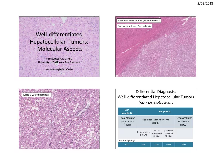
Nancy Joseph, MD, PhD University of California, San Francisco Nancy.Joseph@ucsf.edu 4 cm liver mass in a 25 year old female Background liver: No cirrhosis What is your differential? Non- neoplastic Neoplastic Focal Nodular Hyperplasia

(FNH)

Hepatocellular Adenoma

(HCA)

Hepatocellular carcinoma

(HCC)

Inflammatory (I-HCA) HNF-1a inactivated (H-HCA) b-catenin activated (B-HCA)

Differential Diagnosis: Well-differentiated Hepatocellular Tumors (non-cirrhotic liver)

Risk of malignancy: None

Low Low

~40% 100%

slide-2
SLIDE 2

5/26/2018 2

Management of benign lesions

FNH

No follow up or treatment necessary

Management of benign lesions

FNH

No follow up or treatment necessary

<5 cm

Resection Annual surveillance

>5 cm b-catenin activated

HCA

Focal nodular hyperplasia (FNH)

  • 30-50 year old women
  • Solitary
  • Asymptomatic
  • Well-circumscribed
  • Central fibrous scar

FNH

slide-3
SLIDE 3

5/26/2018 3

FNH

Useful Immunohistochemistry in FNH: Glutamine synthetase (GS) Normal liver FNH

  • Enzyme involved in

nitrogen metabolism expressed in centrizonal hepatocytes H&E

Diagnostic challenge in FNH: Small biopsy

Glutamine synthetase (GS)

Focal nodular hyperplasia: Molecular Aspects

  • No recurrent copy number alterations or

mutations identified

– Multiple prior studies have examined molecular feautures of FNH – Capture based next generation sequencing (500 cancer genes) – Gene expression profiles show overexpression of the b-catenin pathway without gene mutation

  • Non-clonal, non-neoplastic
  • No metastasis of FNH have ever been reported

Rebouissou et al. Hepatology 2008 (49)

slide-4
SLIDE 4

5/26/2018 4

Risk of malignancy: None

Low Low

40% 100%

Non- neoplastic Neoplastic Focal Nodular Hyperplasia

(FNH)

Hepatocellular Adenoma

(HCA)

Hepatocellular carcinoma

(HCC)

Inflammatory (I-HCA) HNF-1a inactivated (H-HCA) b-catenin activated (B-HCA)

Classification of Hepatocellular Tumors Inflammatory HCA: Clinical features

  • ~40-50% of all HCA
  • Female gender
  • Associated with obesity and metabolic

syndrome

– steatosis or steatohepatitis can be present in the non-neoplastic liver

Inflammatory HCA Reticulin-Inflammatory HCA

slide-5
SLIDE 5

5/26/2018 5

Inflammatory HCA

Histologic features FNH Inflammatory HCA

Nodular architecture 85% 7% Fibrous bands 90% 26% Ductular reaction 83% 43% Inflammation 48% 60% Sinusoidal dilatation 18% 83%

Diagnostic challenge: Histologic overlap with Inflammatory HCA

Joseph/Kakar, Mod Pathol 2014 (27)

C Reactive Protein (CRP) Serum amyloid A (SAA)

Useful Immunohistochemisty in I-HCA

Genetic alterations in Inflammatory HCA

Pilati et al. Cytokine & Growth Factor Reviews 2015; 26:499-506

IL6ST

slide-6
SLIDE 6

5/26/2018 6

Genetic alterations in Inflammatory HCA

Pilati et al. Cancer Cell 2014

HNF1-a inactivated HCA: Clinical features

  • Hepatocyte nuclear factor 1a
  • ~30-40% of HCA
  • Female gender
  • Can have multiple tumors/adenomatosis

HNF1-a inactivated HCA

Useful Immunohistochemistry in H-HCA:

Loss of liver fatty acid binding protein (LFABP)

slide-7
SLIDE 7

5/26/2018 7

  • LFABP staining can be lost in HCC
  • Reticulin often lost in fatty areas

Diagnostic challenge in H-HCA: Differentiating from HCC

Genetic alterations HNF1-a inactivated HCA

Pilati et al. Cancer Cell 2014

b-catenin activated HCA (B-HCA):

Clinical Features

  • Male gender (40%)
  • Age > 50 years
  • Androgen use
  • Usually a solitary lesion
  • Glycogen storage diseases
  • Risk for HCC

H&E Reticulin

b-catenin activated HCA

~40% of cases show cytologic

  • r architectural atypia
slide-8
SLIDE 8

5/26/2018 8

b-catenin (nuclear) Glutamine Synthetase (GS)

b-catenin activated HCA (B-HCA):

Useful immunohistochemistry

Diffuse homogeneous GS 90-100% cells Diffuse heterogeneous GS 50%-90% cells Patchy GS < 50%

Hale/Kakar, Mod Pathol 2016

b-catenin activated

b-HCA Diagnostic Challenge:

Glutamine Synthetase staining can be difficult to interpret

β-catenin activated HCA: Genetic Alterations

WNT/b-catenin signaling pathway

– APC/AXIN/GSK3 are negative regulators of this pathway – GLUL (Glutamine Synthetase) is a downstream gene that is upregulated with b-catenin activation – Frequent Copy Number Changes (similar to those seen in HCC)

Zucman-Rossi, Hepatol 2006 Bioulac-Sage, Hepatol 2007 Evason/Kakar, Hum Pathol 2013

b-HCA Molecular Profile: Comparison of cases with and without cytoarchitectural atypia

b-HCA

p value No Atypia (n=5) Atypia (n=6) Copy Number Alterations 1/5 (20%) 3/6 (50%) 0.35 ≥ 1 additional pathogenic molecular alteration 2/5 (40%) 3/6 (50%) 0.43

Joseph/Kakar, Unpublished data

slide-9
SLIDE 9

5/26/2018 9

Well-differentiated HCC with diffuse GS

H&E Reticulin Glutamine synthetase

b-HCA (n=11) Well-diff HCC (n=7) p-value

WNT pathway mutations

8/11 (73%) 5/7 (72%) <0.68

TERT promoter mutation

0/11 (0%) 3/7 (43%) 0.04

≥ 1 additional pathogenic molecular alteration

5/11 (45%) 6/7 (86%) 0.10

b-HCA Molecular Profile: Comparison to Well-diff HCC with diffuse GS

Joseph/Kakar, Unpublished data

Frequency of copy number changes in HCA

Pilati et al. Cancer Cell 2014

p

Non- neoplastic Neoplastic Focal Nodular Hyperplasia

(FNH)

Hepatocellular Adenoma

(HCA)

Hepatocellular carcinoma

(HCC)

Inflammatory (I-HCA) HNF-1a inactivated (H-HCA) b-catenin activated (B-HCA)

None

Low Low

40% 100%

Classification of Hepatocellular Tumors

Risk of malignancy:

slide-10
SLIDE 10

5/26/2018 10

Non- neoplastic Neoplastic Focal Nodular Hyperplasia

(FNH)

Hepatocellular Adenoma

(HCA)

Atypical Hepatocellular carcinoma

(HCC)

Inflammatory (I-HCA) HNF-1a inactivated (H-HCA) b-catenin activated (B-HCA)

Risk of malignancy: None

Low Low

40% 100%

Classification of Hepatocellular Tumors

4 cm liver mass in a 25 year old female Case #2 – 30 M with liver mass

Liver fatty acid binding protein (LFABP)

4 cm liver mass in a 25 year old woman

CRP SAA

Case #2 – 30 M with liver mass Case #1 – 4 cm mass in a 25 year old woman Any other stains?

slide-11
SLIDE 11

5/26/2018 11

Reticulin

4 cm liver mass in a 25 year old woman Any other stains? Case #2 – 30 M with liver mass

Glutamine synthetase

Case #1 – 4 cm mass in a 25 year old woman

Genetic alterations in 250 HCA

Pilati et al. Cancer Cell 2014

Final diagnosis: Atypical hepatocellular neoplasm; see comment.

  • Other terminologies

– Hepatocellular neoplasm with uncertain malignant potential – Well-differentiated hepatocellular neoplasm with atypical or borderline features – Inflammatory adenoma with b-catenin activation

slide-12
SLIDE 12

5/26/2018 12

Take home points

  • Use a panel for well-differentiated lesions

– Glutamine Synthetase (and b-catenin) – Reticulin – CRP or SAA – LFABP

  • Important to identify b-catenin activation
  • Glutamine synthetase stain is much more sensitive

than b-catenin stain, but can be challenging to interpret in some cases

  • CTNNB1 sequencing

Nault et al. J Hepatology 2017 Nault et al. J Hepatology 2017 Nault et al. J Hepatology 2017

slide-13
SLIDE 13

5/26/2018 13

34 year old woman, liver nodule biopsied Glutamine synthetase 34 year old woman, died of drug abuse, liver nodule biopsied LFABP

H&E Reticulin

Diagnosis? HNF1a inactivated adenoma? b-catenin activated adenoma? HCC?

slide-14
SLIDE 14

5/26/2018 14

34 year old woman, died of drug abuse, liver nodule biopsied HMB-45 Arginase

  • Perivascular epithelioid cell tumor (PEComa)
  • Variable admixure of spindled cells, adipose, and

thick-walled vessels

  • Most commonly seen in the kidney
  • Associated with Tuberous Sclerosis (germline and

somatic)

  • Hepatic AML are usually sporadic

– 5-15% are syndromic

  • Hepatic AML commonly show predominant

epithelioid component

Angiomyolipoma

UCSF GI-Liver Pathology