Intrahepatic cholangiocarcinoma Histologic spectrum, novel markers - - PDF document

intrahepatic cholangiocarcinoma
SMART_READER_LITE
LIVE PREVIEW

Intrahepatic cholangiocarcinoma Histologic spectrum, novel markers - - PDF document

5/7/2018 2018 Current Issues in Surgical Pathology Summary (not actual lecture) Intrahepatic cholangiocarcinoma Histologic spectrum, novel markers and molecular assays Sanjay Kakar, MD University of California, San Francisco Intrahepatic


slide-1
SLIDE 1

5/7/2018 1

Intrahepatic cholangiocarcinoma

Histologic spectrum, novel markers and molecular assays

Sanjay Kakar, MD University of California, San Francisco

2018 Current Issues in Surgical Pathology Summary (not actual lecture)

Intrahepatic cholangiocarcinoma

  • Molecular alterations and

histologic classification

  • Diagnostic challenges

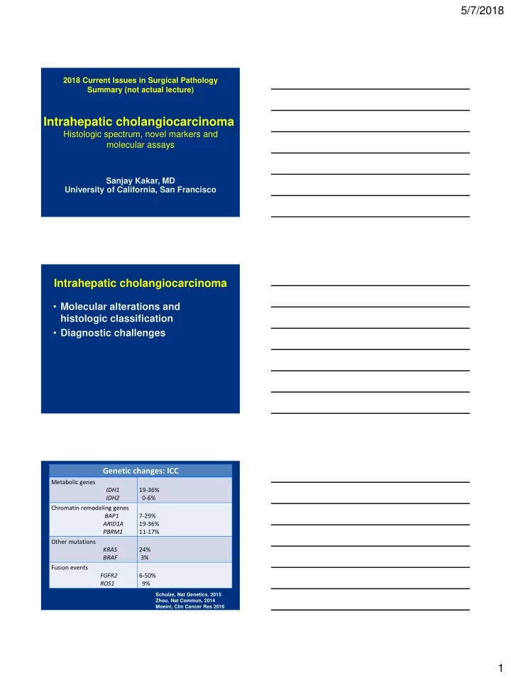
Schulze, Nat Genetics, 2015 Zhou, Nat Commun, 2014 Moeini, Clin Cancer Res 2016

Genetic changes: ICC

Metabolic genes IDH1 IDH2 19-36% 0-6% Chromatin remodeling genes BAP1 ARID1A PBRM1 7-29% 19-36% 11-17% Other mutations KRAS BRAF 24% 3% Fusion events FGFR2 ROS1 6-50% 9%

slide-2
SLIDE 2

5/7/2018 2

ICC vs. HCC

Genetic change Hepatocellular carcinoma Intrahepatic cholangiocarcinoma

β-catenin mutation TERT promoter mutation 20-30% 30-50% Uncommon Rare IDH mutations PBRM1 mutation FGFR2 fusion Rare Absent Absent 19-36% 6-50%

ICC vs. metastatic adenocarcinoma

Genetic change ICC Biliary AC GB PDAC Eso/Gastric IDH mutations 19-36% 0-7% BAP1 mutation 7-29% 0-10% <1% 3% PBRM1 mutation 11-17% 5% 20% 4-6% SMAD4 mutation 0-4% 10-25% 35-60% 8% FGFR2 fusion 6-50% 0-5% 20% 2-9%

Intrahepatic cholangiocarcinoma

Histologic classification

Adenocarcinoma Well-differentiated Moderately-differentiated Poorly-differentiated Histologic subtypes Mucinous Signet ring Clear cell Lymphoepithelioma-like Sarcomatoid Adenosquamous Squamous

slide-3
SLIDE 3

5/7/2018 3 Intrahepatic cholangiocarcinoma

Proposed classification #1 Classification based on size of glands

Large duct type (Mucin-ICC) Located close to hilum Glands typically large Mucin-positive Small duct-type (Mixed-ICC) Located at periphery Glands typically small Mucin-negative Cholangiolocellular Ductular reaction-like pattern

Komuta, Hepatol 2012

Intrahepatic cholangiocarcinoma

Proposed classification #2 Classification #2

Conventional ICC With biliary features Unconventional ICC Trabecular subtype Hilar subtype With predominant ductal plate malformation Cholangiolocellular Intraductal neoplasia Intraductal papillary neoplasm Intraductal tubulopapillary neoplasm

Sempoux, Sem Liv Dis 2011

Intrahepatic cholangiocarcinoma

  • Molecular alterations and

histologic classification

  • Diagnostic challenges
slide-4
SLIDE 4

5/7/2018 4 Intrahepatic cholangiocarcinoma

Diagnostic challenges

Well-differentiated

  • BDA vs adenocarcinoma

Adenocarcinoma: any differentiation

  • ICC vs metastatic adenocarcinoma

Poorly differentiated

  • Hepatocellular carcinoma

Well-differentiated

Frozen section diagnosis

  • Metastatic adenocarcinoma
  • Benign biliary proliferation: Bile duct

adenoma or hamartoma

Benign biliary lesions

Biliary hamartoma (von Meyenburg complex)

  • Dilated ducts with curvilinear outlines
  • Inspissated bile

Bile duct adenoma

  • Compact small to medium sized glands
  • Round to oval contours, not dilated
  • Scant stroma if small, larger lesions can have

prominent stroma

slide-5
SLIDE 5

5/7/2018 5

Biliary hamartoma Bile duct adenoma

Glands Dilated ducts with curvilinear outlines Compact small to medium sized glands Stroma Can be prominent Scant in small lesions, can be prominent in large lesions Bile or eosinophilic material Often present Absent

Bile duct adenoma Adenocarcinoma

Growth pattern

  • Well-demarcated at

interface

  • Grow around portal

tracts

  • Destructive growth
  • Portal invasion

Stroma Typically collagenized in center, can be cellular Desmoplastic Cytologic atypia Mild Mild to marked Architecture Tubular glands, can be angulated Can be complex Mitoses Absent Can be present

Bile duct adenoma

Challenging features

  • Atypia enhanced by frozen artifact
  • Angulated infiltrative glands
  • Mucin can be present
  • Stroma may simulate desmoplasia
  • Variant histologic features
slide-6
SLIDE 6

5/7/2018 6 Bile duct adenoma

Variant features

  • Clear cell change
  • Oncocytic change
  • Alpha-1-antitrypsin globules
  • Granulomas

Bile duct adenoma

Benign neoplasm or reactive process

  • Reactive proliferation related to prior

injury

  • Peribiliary gland hamartoma
  • Benign neoplasm

Bile duct adenoma

Study Result

Pujals, Hepatology 2015 53% had BRAF V600E mutation Pujals, Histopathol 2015 53% positive for VE1 antibody by immunohistochemistry Angkathunyakul, Histopathol 2017 87.5% had BRAF V600E mutation

slide-7
SLIDE 7

5/7/2018 7

BRAF mutation in ICC

Study Result

Goeppert, Mod Pathol 2014 ICC: 5/159 (3%) Extrahepatic biliary, GB: negative Zhu, Ann Surg Oncol 2014 ICC: 4.9% Lee, JCP 2016 Extrahepatic biliary: 1%

BDA vs adenocarcinoma

Immunohistochemistry: p53, Ki-67

IHC Result

p53 Strong diffuse staining 35% ICC 60% metastatic PDAC None: bile duct adenoma (patchy weak to moderate staining) Ki-67 index >10% ICC: 88.5% (mean >20%) BDA: none (mean 2%)

Tan, AIMM 2004 Hornick, AJSP 2005 Tsokos/Gill, Histopathol 2016

BDA vs adenocarcinoma

Immunohistochemistry: Other assays

IHC Result

DPC4 loss ICC: 5-10%; metastatic PDAC 50-60% BDA: none Mesothelin Metastatic PDAC: 64% BDA: none mCEA Metastatic PDAC: 92% BDA: none BAP1 loss ICC: 20-30% BDA: none (limited experience) Albumin ISH Metastatic PDAC: none BDA: Positive

Hornick, AJSP 2005 Arora, Histopathol 2016 Misumi, Histopathol 2017

slide-8
SLIDE 8

5/7/2018 8

Cholangiolocellular carcinoma

Histologic criteria for diagnosis

Study Microscopic Description Inconsistencies Kozaka, 2007

  • small tubular or acinar or

cord-like structures, resembling reactive bile ductules Some studies:

  • Small tubular glands enough

for diagnosis, did not require branching configuration Some studies:

  • CCC divided into well,

moderate and poorly differentiated categories Komuta, 2008 • small monotonous glands

  • antler-like anastomosing

patterns

  • abundant hyalinized and/or

edematous fibrous stroma Moeini, 2017

  • glands strongly embedded

in fibrous stroma Rhee, 2018

  • cuboidal to low columnar

cells with scanty eosinophilic

  • r amphophilic cytoplasm
  • small monotonous glands

CCC: ‘stem cell’ features?

Study Stem Cell Markers

Kozaka, 2007

  • NCAM (CD56): 75%

Komuta, 2008

  • NCAM in 87%
  • CD133, c-kit, OCT4

Moeini, 2017

  • SALL4: 75%
  • NCAM:100%

Rhee, 2018

  • No difference in stem cell marker staining

compared to intrahepatic cholangioCA

Cholangiolocellular carcinoma

slide-9
SLIDE 9

5/7/2018 9

Cholangiolocellular carcinoma Well-differentiated ICC

CD56 in CCC

positive and negative case

EMA in CCC

Luminal and cytoplasmic

EMA per literature

  • Luminal

staining in CCC

  • Cytoplasmic

staining in ICC

Komuta, Hepatol 2012 Kondo, Int Med 2015

slide-10
SLIDE 10

5/7/2018 10

Adenocarcinoma

Any differentiation ICC vs metastatic adenocarcinoma

  • Pancreas, biliary tree
  • Upper GI

ICC vs PDAC

Immunohistochemistry: not widely studied

Diagnosis IHC results

ICC S100P- pVHL+ MUC5AC- CK17- PDAC S100P+ pVHL- MUC5AC+ CK17+

Lok, Hum Pathol 2014

ICC vs PDAC

Other assays

Diagnosis IHC results

ICC BAP1 loss Albumin ISH positive PDAC BAP1 intact Albumin ISH negative

slide-11
SLIDE 11

5/7/2018 11

BAP1: loss in tumor cells BAP1 loss

Poorly differentiated ICC vs HCC

  • ICC with solid/trabecular growth

pattern

  • Scirrhous HCC
  • Combined HCC-cholangiocarcinoma
slide-12
SLIDE 12

5/7/2018 12

Cholangiocarcinoma HCC-like area HCC-like area CK19+ (Arg neg) Scirrhous HCC

slide-13
SLIDE 13

5/7/2018 13

Hep Par, pCEA MOC31

Scirrhous HCC: Atypical features

  • Atypical radiologic features
  • Abundant stroma
  • Immunophenotypic features

Negative: Hep Par 1, pCEA Positive: MOC31, CK19 GPC-3 CK19

slide-14
SLIDE 14

5/7/2018 14

Stain Scirrhous HCC Conventional HCC

Hep Par 1 17-20% 80-90% pCEA 33% 60-80% CK7 58-65% 0-20% CK19 50% 0-10% MOC31 64% 5-11%

Matsuura, Histopath, 2005 Krings/Kakar, Mod Pathol 2013

Arginase-1 95% 95% Glypican-3 95% 70-80%

Combined HCC-CC

HCC

  • Morphology, arginase-1
  • CK19: can be positive

CC

  • Discrete glands, mucin +
  • Negative arginase-1
  • CK7, CK19 and/or MOC31

HCC-like area Well-formed glands

slide-15
SLIDE 15

5/7/2018 15

Arginase-1 CK19 Arginase-1 CK19 Intrahepatic cholangiocarcinoma

AJCC 8th edition

T category Definition

T1 T1a: Solitary tumor <5 cm without vascular invasion T1b: Solitary tumor >5 cm without vascular invasion T2 Solitary tumor with intrahepatic vascular invasion,

  • r multiple tumors, with or without vascular

invasion T3 Tumor perforates visceral peritoneum T4 Tumor involving local extrahepatic structures by direct invasion

slide-16
SLIDE 16

5/7/2018 16

Intrahepatic cholangiocarcinoma

AJCC 7th edition

T category Definition

T1 Solitary tumor without vascular invasion T2 T2a: Solitary with vascular invasion T2b: Multiple tumors T3 Involving visceral peritoneum or direct invasion into extrahepatic structures T4 Tumor with periductal invasion

Periductal invasion

  • Intrahepatic CC, macroscopic types

Mass forming, periductal, intraductal, mixed

  • Periductal: worse prognosis

Extensive intraductal growth: T4

  • Problems

How extensive is 'extensive' Recent studies do not confirm worse outcome

Hirohashi, Hepatogastroeterol 2002 Uno, Surg Today, 2012

Periductal growth pattern

slide-17
SLIDE 17

5/7/2018 17

Intrahepatic cholangiocarcinoma, 3 cm, no VI

Summary

  • Mutations in IDH1, PBRM1 and BAP1, and

FGFR2 fusion can help in distinction of ICC from metastatic adenocarcinoma

  • Mutation in IDH1 and PBRM1, and FGFR2

fusion along with absence of CTNNB1 and TERT promoter mutation helps in distinction from HCC

Summary

BAP1 loss by IHC

  • Can help in distinction from PDAC and upper

GI metastatic adenocarcinoma

  • Can help in distinction from benign biliary

lesions

slide-18
SLIDE 18

5/7/2018 18

Summary

p53 and Ki-67 IHC

  • Diffuse strong p53 staining supports

adenocarcinoma over bile duct adenoma

  • Ki-67 proliferation index >10% is rare in bile

duct adenoma

Summary

HCC vs ICC

  • Diagnosis has strong impact on treatment
  • Sensitive markers like arginase-1 should be

used to identify HCC component

  • ICC diagnosis should be based on stric t

criteria: discrete glands, mucin+, positive for CK19/CK7, typically negative for hepatocellular markers