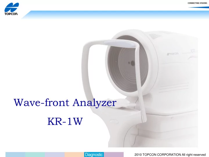

Wave-front Analyzer KR-1W 2010 TOPCON CORPORATION All right reserved
Measure Refractometory Keratometry Aberrometry Topography Pupillometry 2010 TOPCON CORPORATION All right reserved
Application - Cataract Cataract Surgery High Quality analysis of Ocular condition. Persuasive tool for the patients. 2010 TOPCON CORPORATION All right reserved
Application- Cornea Corneal Diseases High Quality analysis of Ocular condition. Helpful information to detect Keratoconus and dry eye patient. Persuasive tool for the patients. 2010 TOPCON CORPORATION All right reserved
Clinical Scene 3 Contact Lens Helpful information for suitable Contact lens selection Helpful information for Dry eye Diagnosis. 2010 TOPCON CORPORATION All right reserved
Clinical Scene 4 Optical Shop Easy screening for ocular abnormality. Helpful information for Night Vision glass prescription 2010 TOPCON CORPORATION All right reserved
Features 5 measurement in one machine Topography with invisible Light Simultaneous Topography and Aberrometry Hartmann-Shack Sensor 10.4 inches Large Touch Panel Display Fully Automated Alignment Multiple Maps for versatile analysis Customizable Screen Layout and Print format 2010 TOPCON CORPORATION All right reserved
Features 10 inches large panel is easier to see. Touch panel monitor enables measuring operation and multiple analysis easier. 2010 TOPCON CORPORATION All right reserved
Features Right and left automated alignment makes it easy for the operator. Just touch the center of the pupil and it automatically measures right and left eyes. 2010 TOPCON CORPORATION All right reserved
Features Fully Automated Alignment and 10.4 inches touch panel display Save tech time with fully automated alignment. Touching the center of the pupil makes automatic start for both right and left eye. 2010 TOPCON CORPORATION All right reserved
Features ; Customizable Display Layout In addition to the regular Maps, preferred custom layout is easily made. (Up to 4 pages) 2010 TOPCON CORPORATION All right reserved
Features; Customizable print format Preferred print format is customizable. (Up to 4 pages) MENU 2010 TOPCON CORPORATION All right reserved
Multiple Maps MENU 2010 TOPCON CORPORATION All right reserved
Multi Maps Find which factor affects the vision, cornea or Crystal lens Ocular Total Aberration Axial Power High Order Aberration Corneal Analysis Ocular Total Analysis 2010 TOPCON CORPORATION All right reserved
Ocular Maps Show the information relating to Ocular maps. Ocular Total Aberration Ocular High Order Aberration RMS ( ㎛ ) Amount of aberration 2010 TOPCON CORPORATION All right reserved
RMS indication per pupil diameters The KR-1W shows RMS data of 4mm (day) and 6mm (night), 4 and 6 mm are statically effective pupil size. RMS ( ㎛ ) Amount of aberration In addition the KR-1W will calculates RMS based on live (actual) pupil size as well! 2010 TOPCON CORPORATION All right reserved
Corneal Maps Indicates the information relating to the cornea. Axial Power Instantaneous map Corneal High Order Aberration RMS ( ㎛ ) Amount of aberration 2010 TOPCON CORPORATION All right reserved
Zernike Vector Maps Find which component of Zernike Polynomials has Ocular total high order aberration. Ocular Total 3rd and 4 th Order aberrations 2010 TOPCON CORPORATION All right reserved
Component Maps Various total ocular, cornea and internal shows where the visual problem exists. Ocular Total Corneal Internal RMS 2010 TOPCON CORPORATION All right reserved
PSF/MTF Maps Patient’s visual simulations. Wavefront-PSF MTF Landolt's’ Ring 2010 TOPCON CORPORATION All right reserved
Valuable information for the refractive Analysis Check the condition at measurement Mire Rings Image Hartman Image Center Shift = Distance between corneal apex and center of pupil. 2010 TOPCON CORPORATION All right reserved
Valuable information for the refractive Analysis RMS and S,C,A is able to compare at the same time. Actual Pupil Size Total High Order Scotopic Photopic Aberration Sphere, Cylinder, Axis data with each pupil diameter 2010 TOPCON CORPORATION All right reserved
Valuable information for the refractive Analysis Wavefront-PSF Normal High Order Aberration These are calculated when the pupil size is 4 mm. 2010 TOPCON CORPORATION All right reserved
Valuable information for the refractive Analysis Contrast sensitivity of patient is visually observed. MTF ( X 、 Y ) MTF (360 ° ) Y X These are calculated when the pupil size is 4 mm. Center Shift (Distance between corneal apex and center of pupil. 2010 TOPCON CORPORATION All right reserved
Valuable information for the refractive Analysis Contrast sensitivity of patient is visually observed. Normal Features High Order Aberration MENU 2010 TOPCON CORPORATION All right reserved
Applications Continuous Measurement MENU 2010 TOPCON CORPORATION All right reserved
Continuous Measurement This measuring function is expected for Dry Eye research. Tear Break Up condition can be observed with 10 times automatic continuous measurement. Features MENU 2010 TOPCON CORPORATION All right reserved
Dry Eye MENU 2010 TOPCON CORPORATION All right reserved
About Dry Eye Dry Eye is depression of chronic tear film stability brought on by single or multiple causes. Destabilization of Corneal epithelium tear film disorder Factors Rack of tear Tear film disappearing supplement inflammation Conjunctiva Infrequent blink Contact lens Depression of Surface wound Corneal aesthesio Surgery Incomplete eyelid close Malfunction of Trichiasis meibomian Features Eye drops MENU 2010 TOPCON CORPORATION All right reserved
Dry Eye Diagnosis Dry Eye Research association 1995 Dry Eye is disease of epithelium brought by quantitative and qualitative abnormality of tear film. ① Shirrmer’s Test : Less than 5mm (5 min.) 2. Abnormity of tear film ② Tear Break-Up time; Less than 5sec. 3. Abnormity on ① Fluorescein stain score ( Cornea + Conjunctiva): epithelium More than 3 points out of 9 points ② Rose-Bengal stain score; More than 3 points out of 9 points All 1,2,3 are abnormal ⇒ Dry Eye 2 out of 1,2 or 3 ⇒ Dry Eye Suspicious MENU 2010 TOPCON CORPORATION All right reserved
Dry Eye Diagnosis Dry Eye Research association 2006 Dry Eye is chronic disease of epithelium and tear film cased by several factors, and it brings on abnormity of visual performance or discomfort of the eye. Changed as below 1.Syndrome abnormity of visual performance or discomfort of the eye ( positive). ① Silmer Test : Less than 5mm (5 min.) 2. Abnormity of tear film ② Tear Break-Up time; Less than 5sec. ① Fluorescein stain score ( Cornea + Conjunctiva): 3. Abnormity on epithelium More than 3 points out of 9 points All 1,2,3 are abnormal ② Rose-Bengal stain score; More than 3 points out of 9 points ⇒ Dry Eye ③ Acid Green Stain Score 2 out of 1,2 or 3 More than 3 points out of 9 points ⇒ Dry Eye Suspicious MENU 2010 TOPCON CORPORATION All right reserved
◆ Shirrmer’s test … For the Dry Eye diagnosis, it is examined to find how much amount of tear is shorter than normal. It is normal when filter paper, hold in bottom lids, hydrated more than 10 mm in 5 minutes, but it is diagnosed to be dry eye when the paper hydrated less than 5 mm. Shirrmer’s test 2010 TOPCON CORPORATION All right reserved
◆ Fluorescein Stain …To see scar existence on the cornea and conjunctiva. ◆ Rose-Bengal Stain …To see condition of the cornea and conjunctiva with the Rose- Bengal Stain. This stains red where mucin layer is not exist and dry, and it reveal where the tear film is dried out in the cornea and conjunctiva. ◆ Acid Green.. To stain transformed epithelium ◆ Fluorescein Stain …To see scar existence on the cornea and conjunctiva. ◆ There are other tests to examine tear film stability or meibomian gland. Fluorescein Stain Rose-Bengal Stain Pictures from Tsurumi Univ Web Site : http://www.tsurumi- eye.com/dryeye/index.html 2010 TOPCON CORPORATION All right reserved
Current Dry Eye examination equipment issue • No instrument is able to evaluate dry eye with new criteria. No product can evaluate visual performance right now. • DR-1 : – The DR-1 shows Oil film BUT ( Break-Up Time), but it is qualitative. – The DR-1 cannot evaluate visual performance. 2010 TOPCON CORPORATION All right reserved
Continuous Measurement The KR-1W would be able to evaluate the dry eye diagnosis method and result with a prescription in terms of visual performance. Tohoku Univ, Prof. Nishida. 2010 TOPCON CORPORATION All right reserved
Study for Dry Eye Stable Dismay Saw Teeth Time Time Time ( sec) ( sec) ( sec) Ocular Total Coma Spherical Shizuka Koh Journal of the eye Vol.24 No.11 Nov.200 MENU 2010 TOPCON CORPORATION All right reserved
Recommend
More recommend