
SLIDE 1
9/30/2016 1
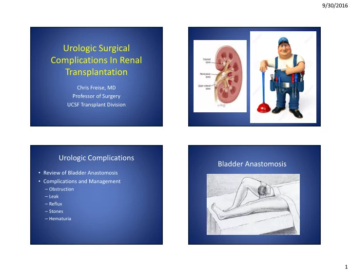
Urologic Surgical Complications In Renal Transplantation
Chris Freise, MD Professor of Surgery UCSF Transplant Division
Urologic Complications
- Review of Bladder Anastomosis
- Complications and Management