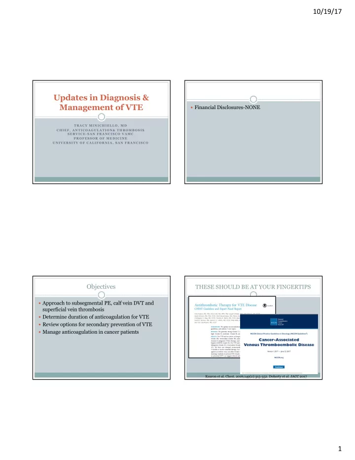

10/19/17 Updates in Diagnosis & Management of VTE Financial Disclosures-NONE TRACY MINICHIELLO, MD CHIEF, ANTICOAGULATION& THROMBOSIS SERVICE- SAN FRANCISCO VAMC PROFESSOR OF MEDICINE UNIVERSITY OF CALIFORNIA, SAN FRANCISCO Objectives THESE SHOULD BE AT YOUR FINGERTIPS Approach to subsegmental PE, calf vein DVT and superficial vein thrombosis Determine duration of anticoagulation for VTE Review options for secondary prevention of VTE Manage anticoagulation in cancer patients Kearon et al. Chest. 2016;149(2):315-352. Doherty et al. JACC 2017 1
10/19/17 Subsegmental PE Volume 41, Issue 1, January 2016 Special Issue: Management of Venous Thromboembolism: Clinical Guidance from the Anticoagulation Forum A 77 yo man had undergoes colectomy for recurrent bleeding from diverticulosis. On POD # 3 he becomes tachycardic to the 110s. WBC is elevated.The surgical team orders an abdominal CT which shows a fluid collection concerning for early abscess. It also shows an isolated RLL subsegmental PE. A dedicated CTa shows a single isolated RLL subsegmenral PE. Do you anticoagulate this patient? Sure, it is a PE. a) No this is incidental. Let’s pretend we don’t know it is b) there Couldn’t you start with an easy question? It is really c) early. Isolated Subsegmental PE Isolated Subsegmental PE Definition: PE shown on CT angiography that occurred in a subsegmental branch but no larger order of vessels. The subsegmental PE may involve one or more than one subsegmental branch Identification of ISSPE has tripled over past decade 2
10/19/17 Isolated Subsegmental PE Subsegmental PE A 77 yo man had undergoes colectomy for recurrent bleeding from diverticulosis. On POD # 3 he becomes tachycardic to the 110s. WBC is elevated.The surgical team orders an abdominal CT which shows a fluid collection concerning for early abscess. It also shows an isolated RLL subsegmental PE. A dedicated CTa shows a single isolated RLL subsegmenral PE. Do you anticoagulate this patient? Sure, it is a PE. a) IS IT REAL? No this is incidental. Lets pretend we don’t know it is b) ISSPE is more likely to be TRUE if….good quality scan, mult defects, centrally there located, d-dimer elevated, seen on mult cuts, patient symptomatic vs Couldn’t you start with an easy question? It is really incidental;high pretest prob of PE c) Get u/s of bilateral lower extrem (upper if CVC) early. Consider risk of recurrence- higher if not post op; immobile; active cancer IF high bleed risk –don’t AC: get serial u/s Kearon et al. Chest. 2016;149(2):315-352. Incidental PE Incidental PE in Cancer A 77 yo man is 2 weeks s/p laproscopic nephrectomy for renal cell CA. He received LMWH for 5 days post op but this was discontinued when he developed melena. An EGD showed a peptic ulcer. He has a staging CT which shows no disease but shows a RUL subsegmental pulmonary artery filling defect. Do you anticoagulated this patient? a) No, that did not go well last time b) Yes, it is a PE c) Easier questions…remember?? 3
10/19/17 Incidental PE in Cancer Incidental PE LOCATION RECOMMENDATION A 77 yo man is 2 weeks s/p laproscopic nephrectomy Proximal DVT or main, lobar AC for at least 6 months for renal cell CA. He received LMWH for 5 days post segmental or multiple op but this was discontinued when he developed subsegmental PE melena. An EGD showed a peptic ulcer. He has a ISSPE with proximal DVT AC for at least 6 months staging CT which shows no disease but does show a RUL subsegmental pulmonary artery filling defect. Do you anticoagulated this patient? ISSPE with distal DVT or no Case be case;consider risk of DVT bleeding/ recurrent thrombosis, a) No, that did not go well last time patient preference. If no b) Yes, it is a PE anticoagulation serial U/S to c) Easier questions…remember?? detect thrombus Calf Vein DVT A 37 year old man presents with right calf pain one week after being kicked in calf during a soccer game. On exam right calf is 2 cm> left. U/S shows DVT in the peroneal vein. What anticoagulation regimen do you recommend? Rivaroxaban 15 mg BID x 21 days then 20 mg daily 1. to complete 3 months of therapy 2. Prophylactic dosing of LMWH or DOAC 3. No anticoagulation, return in one week for repeat ultrasound of lower extremity. 4. Um, is that a deep vein? The guy sitting next to me wants to know. Also includes gastroc and soleus veins 4
10/19/17 Calf Vein DVT- CHEST 2016 Calf Vein DVT -CHEST 2016 AC Forum clinical guidance We suggest treatment of distal DVT with anticoagulation versus observation. We suggest a duration of therapy 3 months. Risk factors for extension: d-dimer +, extensive thrombosis close to proximal veins; active cancer, prior VTE, inpatient Streiff MB et al. J Thromb Thrombolysis . 2016;41:32-67. . Kearon et al. Chest. 2016;149(2):315-352. Calf Vein DVT Calf Vein DVT A 37 year old man presents with right calf pain on week after being kicked in calf during a soccer game. On exam right calf is 2 cm> left. U/S shows DVT in the peroneal vein. What anticoagulation regimen do you recommend? Rivaroxaban 15 mg BID x 21 days then 20 mg daily 1. to complete 3 months of therapy 2. Prophylactic dosing of LMWH or DOAC • 1 st DVT, no cancer, outpatient only 3. No anticoagulation, return in one week for repeat • 6 weeks LMWH and GCS vs placebo and GCS ultrasound of lower extremity. • U/S at 3-7 days and 42 days 4. Um, is that a deep vein? The guy sitting next to me • Outcome progression to proximal DVT or PE wants to know. • No difference in VTE, increased risk of bleeding Righini et al. Lancet Haematol 2016;3: e556–62 5
10/19/17 Superficial Vein Thrombosis Superficial Vein Thrombosis –CHEST Guidelines A 55 year old woman presents with painful swelling Factors that favor the use of AC : extensive SVT; over anterior left thigh. On exam she has a palpable above the knee, close to saphenofemoral junction; cord concerning for SVT. She has an u/s which shows severe symptoms; involvement of the greater saphenous vein; history of VTE or SVT; active thrombosis of the greater saphenous vein extending cancer; recent surgery from the calf proximally and terminating 6 cm from In patients with superficial vein thrombosis of the the deep femoral vein. What do you recommend? lower limb of at least 5 cm in length, we suggest the Prophylactic fondaparinux a. use of a prophylactic dose of fondaparinux or LMWH for 45 days over no anticoagulation (Grade b. Prophylactic rivaroxaban 2B). Full dose DOAC or warfarin c. d. NSAIDS and ice CALISTO TRIAL- fonda vs placebo Primary outcome 1% vs 6% Kearon C et al. Chest . 2012 Superficial Vein Thrombosis Superficial Vein Thrombosis • >400 pts symptomatic SVT riva 10 mg v fonda 2.5mg • Symptomatic above the knee SVT of at least ≥ 5 cm length + other risk factor (>65 , male,hx VTE , cancer, autoimmune disease, non-varicose veins) • No difference in primary efficacy outcome Full dose anticoagulation • After 6 weeks 7% recurrence risk in high risk patients for at LEAST 6 weeks (v 1.2% in CALISTO) 6
10/19/17 Superficial Vein Thrombosis Duration of Anticoagulation for VTE A 55 year old woman presents with painful palpable A 57 year old man presents with unprovoked PE. He swelling over anterior left thigh. On exam she has a has no other PMHx. He is started on rivaroxaban. palpable cord concerning for SVT. She has an u/s How long should he remain on anticoagulation? which shows thrombosis of the greater saphenous vein 1) One year extending from the calf proximally and terminating 2 cm from the deep femoral vein. What anticoagulant 2) 6 months regimen do you recommend? 3) 3 months Prophylactic fondaparinux a. 4) Indefinitely b. Prophylactic rivaroxaban 5) At least until I sign out Full dose DOAC or warfarin c. d. Nsaids and ice Risk of VTE Recurrence After Risk of VTE Recurrence After AC Is Stopped Anticoagulation Is Stopped Characteristic Recurrence at 1 y Recurrence at 5 y Other Helpful Tools Independent Predictors of VTE Recurrence 1,2 Major provoked 1% 3% Increasing patient age Age- and sex-adjusted D- (transient) dimer cutoff levels 3 Increasing BMI Minor provoked 5% 15% Clinical prediction tools 4 (transient) Male gender Unprovoked 10% 30% ¡ DASH Active cancer Cancer 20% — ¡ Vienna Second episode of ¡ Men Continue and HER- unprovoked VTE DOO2 Major transient risk factors + D-dimer after stopping Nontransient risk Major surgery, trauma factors anticoagulation Minor transient risk factors Active cancer, severe PE higher risk for recurrent Pregnancy, minor surgery, long- thrombophilia, inflammatory PE haul air travel, immobilization bowel disease 1. Kearon C et al. Blood . 2014;123:12. 2. Heit JA. Nat Rev Cardiol . 2015;12(8):464-474. 3. Palareti G et al. Int J Lab Kearon C et al. Blood . 2014;123(12):1794-1801 . Hematol . 2016;38(1):42-49. 4. Kyrle PA et al. Thromb Haemost . 2012;108:1061-1064. 28 27 2. Heit JA. Nat Rev Cardiol . 2015;12(8):464-474. 7
Recommend
More recommend