
SLIDE 1
Unit IV – Problem 1 – Clinical: Clinical Presentation of Type-I Diabetes Mellitus
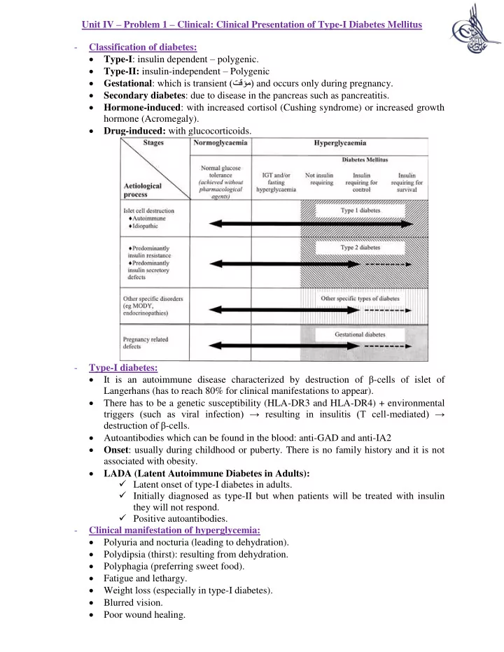
- Classification of diabetes:
Type-I: insulin dependent – polygenic. Type-II: insulin-independent – Polygenic Gestational: which is transient (تقؤم) and occurs only during pregnancy. Secondary diabetes: due to disease in the pancreas such as pancreatitis. Hormone-induced: with increased cortisol (Cushing syndrome) or increased growth hormone (Acromegaly). Drug-induced: with glucocorticoids.
- Type-I diabetes:
It is an autoimmune disease characterized by destruction of β-cells of islet of Langerhans (has to reach 80% for clinical manifestations to appear). There has to be a genetic susceptibility (HLA-DR3 and HLA-DR4) + environmental triggers (such as viral infection) → resulting in insulitis (T cell-mediated) → destruction of β-cells. Autoantibodies which can be found in the blood: anti-GAD and anti-IA2 Onset: usually during childhood or puberty. There is no family history and it is not associated with obesity. LADA (Latent Autoimmune Diabetes in Adults): Latent onset of type-I diabetes in adults. Initially diagnosed as type-II but when patients will be treated with insulin they will not respond. Positive autoantibodies.
- Clinical manifestation of hyperglycemia: