D complaint, with infection, irritable bowel syndrome, infmammatory - - PDF document

d
SMART_READER_LITE
LIVE PREVIEW

D complaint, with infection, irritable bowel syndrome, infmammatory - - PDF document

PRACTICALGASTROENTEROLOGY SEPTEMBER2018 A CASE REPORT Autoimmune Enteropathy: An Uncommon Presentation by Alan Naim, Kaivan Salehpour, Camron Kiafar INTRODUCTION iarrhea is a common gastrointestinal D complaint, with


slide-1
SLIDE 1

70฀

PRACTICAL฀GASTROENTEROLOGY฀ •฀ SEPTEMBER฀2018

A CASE REPORT

Autoimmune Enteropathy: An Uncommon Presentation

Alan Naim MD,1,3 Kaivan Salehpour MD,2 Camron Kiafar DO, AGAF, FACG1,3 1Department

  • f

Gastroenterology, University

  • f

Arizona College

  • f

Medicine-Phoenix, Phoenix, AZ

2Department of Internal Medicine, University

  • f

Arizona College

  • f

Medicine-Phoenix, Phoenix, AZ 3Department of Gastroenterology, Phoenix VA Health Care System, Phoenix, AZ

by Alan Naim, Kaivan Salehpour, Camron Kiafar colonoscopy at the outside hospital was normal. However, random biopsies were suggestive of lymphocytic colitis for which the patient was discharged on budesonide therapy along with total parenteral nutrition (TPN). He presented to our hospital with ongoing intractable diarrhea despite compliance to his medication therapy. At the time of presentation, he endorsed greater than 15 watery stools per day with nocturnal

  • symptoms. A gluten-free and lactose-free diet

failed to improve his symptoms. He had lost nearly 20 pounds in the last two months. He denied recent travel or sick contacts. He had no history of autoimmune disease as well as no family history

  • f gastrointestinal or autoimmune disease. He had

a longstanding smoking history but denied alcohol

  • r drug use.

Vital signs were all within normal limits. On physical exam, his fjndings were only signifjcant for a deconditioned gentleman as well as rectal exam showing peri-anal excoriations. Laboratory studies demonstrated a normocytic anemia, elevated INTRODUCTION

D

iarrhea is a common gastrointestinal complaint, with infection, irritable bowel syndrome, infmammatory bowel disease (IBD) and malabsorption syndromes (such as lactose intolerance and celiac disease) being the most common etiologies. We report a case

  • f a 70-year old male with a two-month history
  • f profuse, watery diarrhea. An abdominal

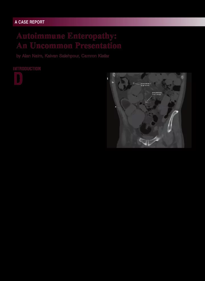
computerized tomography (CT) scan on initial presentation revealed pneumatosis intestinalis. Extensive workup that included enteroscopy and colonoscopy revealed histology suggestive

  • f autoimmune enteropathy. Anti-enterocyte

antibodies confjrmed the diagnosis. Although rare, autoimmune enteropathy (AIE) represents an important consideration in the differential diagnosis of intractable diarrhea in adults.

Presentation

A 70-year old male with a history of resected Meckel’s diverticulum and acid refmux was admitted for profuse, frequent, watery diarrhea and

  • hypotension. Two months prior, he had presented

to an outside hospital with similar symptoms including mild abdominal pain; he was found to be severely dehydrated with associated acute kidney

  • injury. At that time, a computed tomography (CT)
  • f the abdomen and pelvis revealed pneumatosis

intestinalis of the small intestine and colon. Due to his presentation and CT fjndings, the patient underwent an exploratory laparotomy. There was no evidence of ischemia, perforation or necrotic bowel noted during laparotomy. Subsequent

Figure 1. CT abdomen/pelvis displaying pneumatosis intestinalis of the small and large intestines.

slide-2
SLIDE 2

A CASE REPORT

PRACTICAL฀GASTROENTEROLOGY฀ •฀ SEPTEMBER฀2018฀

71 Autoimmune Enteropathy: An Uncommon Presentation again demonstrated pneumonitis intestinalis of the small intestine and colon (Figure 1). Given his non-response to budesonide therapy, and in consideration for an alternative diagnosis, the decision was made to undergo upper enteroscopy and repeat colonoscopy. Esophagogastroduodenoscopy (EGD) and upper enteroscopy fjndings were normal and random biopsies of the duodenum and jejunum were

  • taken. The colonoscopy showed boggy, edematous

appearing mucosa. Random biopsies of the colon were taken as well. Small bowel biopsies all indicated moderate to severe villous blunting, with marked lamina propria infjltrate (plasma cells and lymphocytes) (Figure 2). No goblet or paneth cells were identifjed. There was marked crypt apoptosis. Colon biopsies exhibited active colitis with preserved crypt

  • architecture. Markedly increased crypt apoptosis

was again demonstrated (Figure 3). No goblet or paneth cells were visualized. Serum Anti-Enterocyte IgG antibodies were present in elevated titers and demonstrated positive linear periapical staining of the enterocytes and staining in the goblet cells. Anti-enterocyte IgA antibody also demonstrated positive linear periapical staining of enterocytes in elevated titers. When the diagnosis of autoimmune enteropathy was established, he was placed on intravenous

  • methylprednisolone. Shortly after, he was started
  • n TPN for nutritional support. The patient began

responding positively to intravenous steroids on the third day of treatment, resulting in a decrease in frequency and volume of bowel movements. As he clinically improved, he was transitioned to oral prednisone and discharged with a steroid taper for

  • utpatient gastroenterology follow-up.

Discussion

Autoimmune enteropathy is a rare cause of intractable diarrhea in children and an even rarer cause in adults. It is best defjned as a presentation of chronic diarrhea, malabsorption, with specifjc small intestinal histologic features and is typically confjrmed by the presence of circulating auto- enteric antibodies. Extraintestinal manifestations may include hypothyroidism, nephrotic syndrome, autoimmune hemolytic anemia, and rheumatoid creatinine, and low albumin level of 2.5 gm/dL (nl 3.4-5.0 gm/dL). Human immunodefjciency virus (HIV) status as well as stool studies, which included stool culture with Shiga toxin, Clostridium diffjcile toxin B PCR, giardia antigen, ova and parasites, fecal lactoferrin and fecal leukocyte testing were all negative. Serum tissue transglutaminase Ab IgA was undetectable with a normal IgA level. CT abdomen/pelvis imaging fjndings at our institution

Figure 2. Jejunal biopsy specimen: findings show dense lamina propria infiltrate comprising of plasma cells and lymphocytes (long arrow). Intraepithelial severe villous blunting is appreciated (short arrow). Figure 2. Colon biopsy specimen: displaying lamina propria infiltrates and crypt apoptosis.

slide-3
SLIDE 3

A CASE REPORT

72฀

PRACTICAL฀GASTROENTEROLOGY฀ •฀ SEPTEMBER฀2018

Autoimmune Enteropathy: An Uncommon Presentation arthritis.1-3 A lack of response to a gluten-free diet

  • r other dietary exclusions is also described, as is

a predisposition to other autoimmune diseases.1-3 The disease was fjrst described in 1982 in London in a 15 month-old child with protracted diarrhea and weight loss. Since that time, only a handful

  • f adult cases of AIE have been reported with the

largest case series from Mayo Clinic Rochester comprising of 15 patients.1 AIE is histologically characterized by fjndings including: villous blunting, increased mononuclear infmammation in the lamina propria, lymphocytic infjltration into deep crypt epithelium with a relative decrease of surface lymphocytosis (< 40 lymphocytes per 100 epithelial cells), as well as the presence of increased crypt apoptotic bodies.1 Mononuclear infjltrates are comprised of both plasma cells and lymphocytes. Colon histopathology shows similar histologic abnormalities to those seen in the small bowel. Gastric biopsies not uncommonly demonstrate an autoimmune atrophic gastritis as well.1 CT fjndings are typically non- diagnostic.1 This is the fjrst known case to report the fjndings of pneumatosis intestinalis in autoimmune enteropathy. The presence of anti-enterocyte or anti-goblet cell antibodies is supportive of the diagnosis of AIE, although the detection of these antibodies has been described as “observer dependent.” The signifjcance of circulating auto-enteric antibodies in regards to pathology has not been fully

  • delineated. Akram et al. did not show association

between the clinical course, intestinal histology, and the type of circulating auto-enteric antibodies.1 While anti-enterocyte antibodies have not been reported in celiac disease and infmammatory bowel disease, anti-goblet antibodies have been reported in patients with chronic infmammatory bowel disease, as well as in their asymptomatic fjrst- degree relatives.4,7,8 Despite concerns regarding their sensitivity and specifjcity, anti-enterocyte antibodies aid in establishing a diagnosis of AIE in cases with protracted diarrhea and malabsorption. Much of the pathophysiology behind autoimmune enteropathy is unknown, but some studies have pointed to a defjciency or dysfunction in CD4+ and CD25+ regulatory T cells which are involved in the down-regulation of a variety

  • f bodily immune responses.5 Mutation in the

FOXP3 gene (forkhead box p3) in T cells, for instance, has been found in inherited forms of autoimmune enteropathy such as the rare X-linked recessive disorder of early childhood known as IPEX (immunodysregulation polyendocrinopathy enteropathy X-linked) syndrome.2,5,6 Scarce data exists regarding epidemiology, disease course, and treatment options for AIE. Anecdotal experience guides a number of treatment decisions including use of corticosteroids as well as immunosuppressive drugs such as azathioprine, cyclophosphamide, tacrolimus, cyclosporine and

  • infmiximab. In a retrospective study, over half of

the patients responded to steroid therapy. However, two-thirds of these patients either became steroid- dependent or refractory requiring additional immunomodulating or biologic therapy for maintenance of remission.1 CONCLUSION Autoimmune enteropathy represents a rare and important consideration in the differential diagnosis of intractable diarrhea in adults. It should be especially sought out in cases of malabsorption and small bowel villous atrophy not responding to a gluten-free diet. This disorder may also be considered in patients presenting with pneumatosis intestinalis when other common causes have been excluded. Treatment can be challenging and often requires both nutritional support with immunosuppressive medications. References

  • 1. Akram S, Murray J, Pardi D, et al. Adult Autoimmune Enteropathy:

Mayo Clinic Rochester Experience. Clin Gastroenterol Hepatol. 2007;5(11):1282-1290.

  • 2. Gentile NM, Murray JA, Pardi DS. Autoimmune Enteropathy: A

Review and Update of Clinical Management. Curr Gastroenterol

  • Rep. 2012;14(5):380-385.
  • 3. Montalto M, D’Onofrio F, Santoro L, et al. Autoimmune enteropathy

in children and adults. Scand J Gastroenterol. 2009;44:1029–1036.

  • 4. Russo PA, Brochu P, Seidman EG, et al. Autoimmune enteropa-
  • thy. Pediatr Dev Pathol. 1999;2:65–71.
  • 5. Yagi H, Nomura T, Nakamura K, et al. Crucial role of FOXP3 in the

development and function of human CD25 CD4 regulatory T cells. Int Immunol. 2004;16(11):1643-1656.

  • 6. Brunkow ME, Jeffery EW, Hjerrild KA, et al. Disruption of a new

forkhead/winged-helix protein, scurfjn, results in the fatal lymphop- roliferative disorder of the scurfy mouse. Nat Genet. 2001;27:68–73.

  • 7. Hibi T, Ohara M, Kobayashi K, et al. Enzyme linked immunosorbent

assay (ELISA) and immunoprecipitation studies on anti-goblet cell antibody using a mucin producing cell line in patients with infmam- matory bowel disease. Gut. 1994;35:224–30.

  • 8. Folwaczny C, Noehl N, Tschop K, et al. Goblet cell autoantibodies

in patients with infmammatory bowel disease and their fjrst-degree

  • relatives. Gastroenterology. 1997;113:101–6.