troponin 35
play

Troponin = 35 ACUTE CORONARY SYNDROME Objectives The first problem - PDF document

2/5/2013 Objectives Improve speed and accuracy in Low Risk assessing patients with possible Chest Pain ACS! Avoid pitfalls in the use of cardiac markers to exclude AMI Jeffrey Tabas, MD Professor of Emergency Medicine Avoid


  1. 2/5/2013 Objectives  Improve speed and accuracy in “Low Risk” assessing patients with possible Chest Pain ACS!  Avoid pitfalls in the use of cardiac markers to exclude AMI Jeffrey Tabas, MD Professor of Emergency Medicine  Avoid pitfalls in the use of non- Office of CME invasive testing to exclude UCSF School of Medicine Unstable Angina 
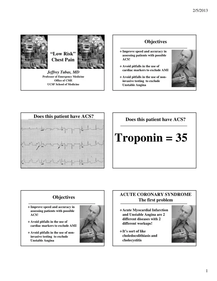
Does this patient have ACS? Does this patient have ACS? Troponin = 35 ACUTE CORONARY SYNDROME Objectives The first problem  Improve speed and accuracy in  Acute Myocardial Infarction assessing patients with possible and Unstable Angina are 2 ACS! different diseases with 2  Avoid pitfalls in the use of different workups! cardiac markers to exclude AMI  It’s sort of like  Avoid pitfalls in the use of non- choledocolithiasis and invasive testing to exclude cholecystitis Unstable Angina 1

  2. 2/5/2013 Case 1 Case 1  54 y.o. M w/ left shoulder ache x 8 Was AMI appropriately excluded? hours. Only hx is smoking ¼ ppd Yes 1.  Normal exam except marked Left No 2. trapezius muscle spasm Care is never appropriate at a conference 3.  ECG – no ischemia lecture  CXR – Normal  Single 8 hr TnI sent What about Unstable Angina? Other diagnoses? = 0.09 [0.00 – 0.10 ng/ml] Case 1 Steps in Assessment of ACS Risk Stratify  Discharged with Dx of shoulder 1. strain and follow-up by PMD Rule out MI 2. in 1-3 days. Rule out UA 3.  Immediate  Patient is brought back 12 hours  Delayed later in cardiac arrest  A lawsuit is brought and settled out of court How do ACS patients How do ACS patients present? - present to our EDs? Summary Gupta, Ann EM 2002 - 720 cases of AMI - 50% of patients with ACS present like the text books say CHEST PAIN CHEST PAIN NO CHEST PAIN (53%) (47%)  50% of patients with ACS present Shortness of Breath (17%) atypically Cardiac arrest (7%) Dizzy/Weak/Syncope (4%)  Atypical is TYPICAL Abdominal Pain (2%) Other (17%) 2

  3. 2/5/2013 Risk Scores TIMI Risk Score TIMI = 0 has sensitivity of 96.6% (91.5-99%)  TIMI - Hess, AEM, 2010  Modified TIMI  GRACE None of the following  FRISC  Age 65 or more  HEART  3 or more CAD risk factors  Known CAD (stenosis >50%)  Most derived from pts with definite ACS, not possible  ASA use in past 7 days ACS (except HEART)  Severe angina (>= 2 episodes w/in 24 hrs)  Based on 1 st troponin - Aren’t we interested after 2nd?  ST changes >= 0.5 mm  Positive initial cardiac marker Simplest Low-risk Score AMI : The Cardiac Markers Risk of events 2% or less  In a patient without ischemia on ECG, it’s all about the troponins!  Negative initial cardiac marker  Near normal ECG  AMI exclusion 6 hours after ONSET is accepted although repeating a level 6 hours after ARRIVAL is common  AMI exclusion 2-3 hours after ARRIVAL is here (but hasn’t reached the guidelines yet) Excluding AMI AMI: ACEP Policy  It’s about the troponins  A negative cardiac marker at least 8 hours from symptom onset  AMI exclusion is something we can and should OR do correctly  A negative 90 min delta myoglobin + (CKMB or ACEP Clinical Policy Troponin) Annals EM, Sept 2006 OR  A negative 2 hr delta CK-MB + Troponin 3

  4. 2/5/2013 What is a Low Risk Patient? Morrow, Circ, 07  No Ischemia on ECG  Initial Cardiac Marker is Negative AMI: Lab Medicine  A negative cardiac marker at least 6 hours from symptom onset IF Low Risk  A negative cardiac marker at least 12 hours from symptom onset IF Mod-High Risk SFGH Protocol for Case 1 ECG and Troponin Testing If symptoms are unchanging or resolved  54 y.o. M w/ left shoulder ache x 8  Check at arrival and at 6 hours from ONSET hours. (not 6 hrs after arrival!)  Nl exam and ECG  E.g. Onset 2hrs prior to arrival, check on arrival and at 4 hrs  Single 8 hr TnI sent  E.g. Onset 6 hrs prior to arrival, check only on arrival = 0.09 [0.00 – 0.10 ng/ml] If symptoms are stuttering  Check on arrival, at 3 hrs, and at 6 hours Was AMI appropriately excluded? Understanding the Lab Understanding the Lab  Assay limit of detection <0 .01  <0.01 = Undetectable  0.01 to 0.1 = Detectable (but within “normal range”)  99 th percentile = 0.10  > 0.1 = Elevated  10% coefficient of variance (imprecision) = 0.3 4

  5. 2/5/2013 Troponin “Leaks” “Acute Troponin Leaks” Aviles and Aviles, EM Clinics, 2005 Newby L, JACC 2012  Tachycardia Saunders JT, Circ 2011  CHF  Any detectable Troponin level is associated with  PE markedly increased adverse event rate over time  Peri/Myocarditis  Renal Failure  DKA  Sepsis Troponin “Leak” – Pearls More rapid ED rule outs? NEJM, Aug 27 2009 Highly sensitive Troponins It’s a leak if: They’ve had it in the past (more than once) 1) You repeat and it doesn’t rise 2) More rapid ED rule outs? Current Troponin Assays - Summary Keller et al, JAMA, Dec 2011  Any detectable level mandates further evaluation - Repeat level and stress testing is safest approach  Although not yet in the guidelines, an  1818 patients in Germany, 23% with AMI undetectable level at 0 and 3 hrs excludes AMI  Highly Sensitive Trop on arrival: Sens = 100% (for level of detection)  Standard Trop 3 hrs post arrival: Sens = 98.2% (for level of detection) 5

  6. 2/5/2013 Super Sensitive Assays - Summary Ways to miss AMI w/ a negative 6 hr Trop Unacceptable  Any detectable level mandates further evaluation  Miss the ischemic ECG - Repeat level and stress testing is safest approach  Troponin not really negative (i.e detectable)  With Highly Senstive Troponins, a 0 and 1 hr  Not really 6 hours after onset (stuttering) level excludes AMI  As sensitivity increases, we will get an Acceptable increasing number of false positives  Very tiny percentage of patients still have AMI  We didn’t miss AMI but unstable angina ED Treadmill Unstable Angina: Noninvasive Tests Amsterdam, JACC, 2002  Understand your non-invasive testing!  1000 ED pts sent for treadmill w/ a single Outpatient testing negative troponin Sensitivity Specificity # of Patients  Negative ETT in 64% = 0.2% Event Rate Treadmill 68 77 24,074  Positive ETT in 13% = 14% Event Rate Nuclear stress 88 77 628 Stress Echo 76 88 1174  Nondiagnostic in 23% = 3.6% Event Rate Lee NEJM 01 Does Prior Stress Testing Exclude ACS? Value of prior stress testing?  “MI evolves most frequently from Nerenberg, AJEM, 07 plaques that are only mildly to Compared with no prior testing: moderately obstructive…the risk of  A positive prior ETT increases admit rate plaque disruption depends more on and rate of adverse events plaque composition and vulnerability  A negative does NOT change admit rate (plaque type) than on degree of stenosis or rate of adverse events (plaque size).” Falk, et al. Circulation, 1995 6

  7. 2/5/2013 Outpatient Noninvasive Testing Lessons from Treadmill testing in 72 hours?  A negative exercise treadmill excludes that  Anderson, Circ, 11 the current symptoms are due to ACS  ACC/AHA Recommends noninvasive testing within 72 hours of ED visit  Confirm that test is diagnostic  85% MPHR (> 6 mets, DP > 22.5 K)  Meyer, Annals EM  Non-diagnostic results need further eval  Showed this was a safe strategy in 1000 low risk Kaiser patients after AMI rule out  Previous treadmill helpful only if abnormal CT Coronary Angio: The Future? Radiation Doses Radiation exposure Yearly background = 3 • CXR = 0.02 • Cardiac cath = 6 • Tc-99 Stress Mibi = 8 • CTCA: Male = 9 mSV (14 if retrospective) • CTCA: Female = 12 (21 if retrospective) • Smith-Bindman, Arch IM 09 - Actual Doses!!!!  CTCA – 22 (14-24) mSv  1000 pts w/o CAD, ischemic ECG or initial positive Tn  Randomized to CTCA or usual care  However, even at 1 year, CTCA vs Usual Care resulted in more tests, more radiation (14 mSv vs 5 mSV), and  2% AMI, 5% UA more interventions (32 vs 21)  Mean LOS reduced by 7.6 hours (P<0.001)  More D/C’s directly from ED: 47% vs. 12% (P<0.001) 7

  8. 2/5/2013 How Can We Detect All ACS? CT in ACS- My Take  The only way to detect all ACS is to test  Excellent Negative Predictive Value everyone!  Use only when treadmill unavailable or  However, we have seen testing lead to wasted patient can’t exercise time, money, unnecessary complications and further testing due to non-diagnostic or false  Probably best for a moderate risk pt positive results i.e. > 10% ACS risk  DO what you and the patient believe is best  Identifies other diseases! (Causes other diseases?) How Do We Manage Our How Can We Detect All ACS? Low Risk Chest Pain Patients?  DOCUMENT their understanding of the risks of the decision, which are always present 8

Download Presentation
Download Policy: The content available on the website is offered to you 'AS IS' for your personal information and use only. It cannot be commercialized, licensed, or distributed on other websites without prior consent from the author. To download a presentation, simply click this link. If you encounter any difficulties during the download process, it's possible that the publisher has removed the file from their server.

Recommend


More recommend