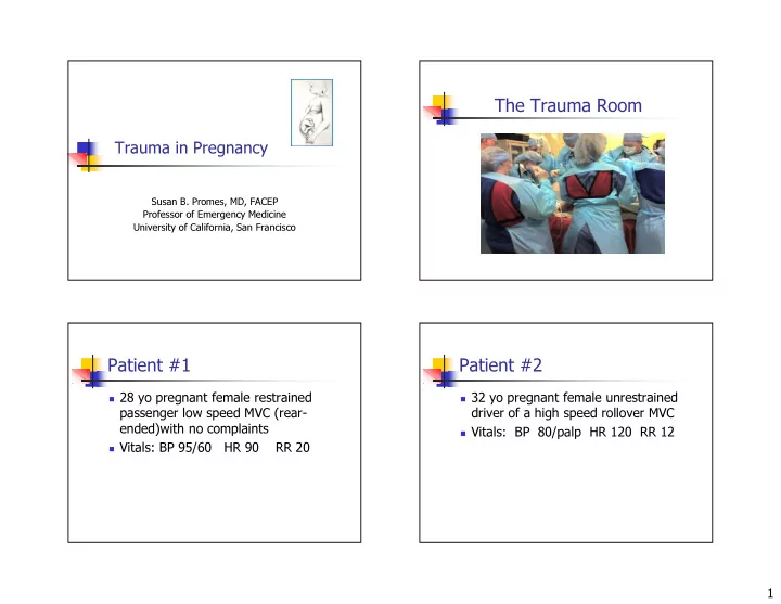

The Trauma Room Trauma in Pregnancy Susan B. Promes, MD, FACEP Professor of Emergency Medicine University of California, San Francisco Patient #1 Patient #2 � 28 yo pregnant female restrained � 32 yo pregnant female unrestrained passenger low speed MVC (rear- driver of a high speed rollover MVC ended)with no complaints � Vitals: BP 80/palp HR 120 RR 12 � Vitals: BP 95/60 HR 90 RR 20 1
Patient #3 Patient #4 � 27 yo pregnant female auto vs pole � 21 yo pregnant female s/p stab wound � Vitals: 160/120 HR 98 RR 24 � Vitals: BP 105/74 HR 100 RR 24 Statistics Outcome � Leading cause of non-obstetric related death clinicians ’ s awareness of altered intra- in pregnant patients Depends on to a great extent the � Occurs in 7-8% of all pregnancies abdominal injury pattern and normal � 2/3 are MVC � 20% related to domestic violence physiologic changes � Prevalence of domestic violence in pregnancy 6- 20% 2
Anatomical Changes Normal Physiologic Changes � Cardiovascular � Respiratory � Hematologic � Gastrointestinal � Metabolic - Endocrine A woman with a normal heart Cardiovascular may have an ECG that appears ischemic. � Cardiac output increases � Pulse rate increases � Blood pressure decreases then returns A. TRUE to baseline B. FALSE � Central venous pressure decreases � ECG changes 3
ECG Changes in Pregnancy Respiratory � Common ECG changes for pregnant � Respiratory rate increases women � Tidal volume increases � LAD � Functional residual capacity decreases � Q wave in III and aVF � Oxygen consumption increases � flattened or inverted T in III � Respiratory alkalosis When would you expect a Hematologic pregnant women’s HCT to be the lowest? � Blood volume increases � Dilutional anemia A. 1 st trimester � WBC count increased B. 2 nd trimester � Platelet count decreased C. 3 rd trimester � ESR increased � Increased risk of thrombolembolic event 4
Lab Values Gastrointestinal Hematocrit (%) Hemoglobin (g/dL) � Motility decreased � Pregnant women: � Pregnant women: � LES tone decreased � 11.4–15.0 � 1st trimester: 35–46 � 10.0–14.3 � 2nd trimester: 30–42 � Albumin and total protein levels � 10.2–14.4 � 3rd trimester: 34–44 decreased � Postpartum: 10.4–18.0 � Postpartum: 30–44 Metabolic-Endocrine Injury Patterns � Total body water increased � Growing uterus effects normal position of other organs � GFR increased � BUN and creatinine decreased � Aldosterone and cortisol levels are increased � Peripheral resistance to insulin 5
Blunt Trauma Penetrating Trauma � MVC - common � If chest tube necessary, consider � Restraints inserting tube higher than usual by a � Location of organs couple rib spaces changed due to � Uterus more prominent pregnancy � Direct fetal injury more likely � Hepatic, splenic, uterine and bladder injuries � GI injuries less common � Think abruption � Can be delayed Pelvic Trauma The Trauma Room � Bony pelvis becomes more lax with pregnancy � Consider repositioning the patient � McRobert or lithotomy � More common injury in pregnancy � Think bowel, bladder and urethral injuries � Vascular injury? 6
Resuscitation � Airway � Breathing � Circulation (positioning key) Manually displace uterus Resuscitation � Airway � Breathing � Circulation (positioning key) � Definitive Treatment ★ Check Rh status IV, oxygen and monitor are key to a successful resuscitation! 7
Radiation Exposure Diagnostics � Abdomen 200-500 mrad � Ultrasound is screening modality of choice � C-Spine < 1 mrad � HOWEVER when US is negative or � Chest 1-3 mrad inconclusive in patient who � L-spine 600-1,000 mrad hemodynamically unstable, DPL may be study of choice � Pelvis 200-500 mrad � Safe in pregnancy � CT brain 1 rad � Use open DPL approach � CT abd/pelvis 1-3 rad Resuscitation ACLS Drugs Category B Category C Category D Atropine Epinephrine Amiodarone Take Care of the Mother First Magnesium Lidocaine Bretylium Bicarbonate Dopamine Dobutamine Adenosine 8
Modifications of CPR � Before fetal viability � No modifications necessary – focus on Hemodynamically mother Stable Patient � After fetal viability (24 weeks) � Patient positioning � Consider C-section Don ’ t forget fetal monitoring! Ultrasound is the test of choice to identify abruption. A. TRUE B. FALSE 9
Placenta Abruptio Placental Abruption � 40-50% major traumas � 1-3% minor traumas � US not sensitive enough � Must monitor � Painful bleeding patients � Blood usually dark � Check Rh status � 20% without bleeding There is no indication to order Kleinhauer Betke test a Kleinhauer Betke test in the ED. Detects transplacental hemorrhage and independent indicator of risk of pre-term labor (LR 20.8) A. TRUE B. FALSE J Trauma. 2004 Nov;57(5):1094-8 10
Monitoring � Fetal heart rate � Variability � Pattern of contractions � Decelerations Uterine Rupture Perimortem C-section � Who? � What? � When? � Why? 11
Who? What to do? � >24 weeks gestation � Get help! � Decide SOON � OB, NICU, Peds, Surg, L&D staff, � Maternal arrest anyone… � preferably sudden � <15 minutes from maternal arrest, � <5 is better, best Effect of Perimortem Perimortem C-section C-section on Maternal Survival Time from RSOC or No change in Arrest improved maternal status Time to GA in # normal total # of hemodynamics Delivery weeks infants infants (min) 0-5 min 25-42 8 11 0-5 5 2 6-10 3 --- 6-10 min 26-37 1 4 11-15 1 --- 11-15 min 38-39 1 2 >15 4 5 >15 min 30-38 4 7 Not reported 1 1 12
Improved Fetal Survival Perimortem C-section � Prognosis best if performed within 5 minutes of maternal arrest and initiation of CPR Fetal age > 28 weeks or 1 kg � � CPR should continue Short interval from maternal death to delivery � Maternal death not from chronic hypoxia during the � Fetal status before maternal death � procedure and brief NICU � time afterward Quality of maternal resuscitation � Perimortem C-section Equipment Critical Steps � Scalpel Continue maternal � resuscitation � Mayo Scissors Vertical midline incision � through abdominal wall � Toothed forceps 4-5 cm below xiphoid to � pubic symphysis � Needle holder Incise fundus � Consider blunt scissors � � Needle and 0 or 1 chromic sutures Deliver baby � APGARS � � Richardson retractors Remove placenta � Oxytocin � 13
Neonatal Life Support ROSC in Mother � Resuscitate � Broad spectrum abx � Carefully close the incision Pregnant Trauma Patients Patient #1 � 27 yo pregnant female restrained passenger low speed MVC (rear- ended)with no complaints � Vitals: BP 95/60 HR 90 RR 20 14
Patient #2 Patient #3 � 32 yo pregnant female unrestrained � 21 yo pregnant female s/p stab wound driver of a high speed rollover MVC � Vitals: BP 105/74 HR 100 RR 24 � Vitals: BP 80/palp HR 120 RR 12 Patient #4 Summary � Must understand � 27 yo pregnant female auto vs pole normal maternal � Vitals: 160/120 HR 98 RR 24 physiology & anatomical changes � Perform perimortem first – but don ’ t C-sections early � Treat the mother forget about the infant 15
Recommend
More recommend