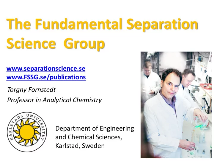

The Fundamental Separation Science Group www.separationscience.se www.FSSG.se/publications Torgny Fornstedt Professor in Analytical Chemistry Department of Engineering and Chemical Sciences, Karlstad, Sweden
Who Are We? Torgny Fornstedt , Emelie Glenne , Prof. Analytical Chemistry PhD-student Jörgen Samuelsson , Joakim Bagge , Assoc. Prof in Surface Biotechnology Researcher Patrik Forssén , Associates: Research Engineer in Scientific Karol Lacki , Computing soon-to-be Adjunct Professor Martin Enmark , Maria Rova , PhD in Chemistry PhD in Biochemistry Marek Lésko , Marek Szymański (Örebro University), Postdoc in Chemical engineering Postdoctoral fellow in Scientific Computing We focus on combining experiments and theory, in order to understand how molecules interact with separation phases and biosensors chips etc.
Our Backgrund at Uppsala University A Surface Biotechnology Center was founded in 2000 to continue the protein separation science at Uppsala University. The Center was sponsored by Amersham Biosciences and professor Karin Caldwell was the leader. Karin Caldwell & Coworkers Jörgen Samuelsson (Right) at the Downstream Proc. Unit 3
Our Site at Karlstad University Jörgen Samuelsson (left) and Marek Leśko (right) in Emelie Glenne and “Wille” 6 the background our SFC instrument Waters UPC2 and months old. furthest away our Waters UHPLC instrument .
Our site at BMC, Uppsala University Dr. Martin Enmark, in the background our Agilent 1200 Instrument.
6 What Are We Doing ? Combining Theory & Practice for Deeper understand how molecules interact with each other and with separation media/biosensor chips Deeper Understanding : Experimental data are processed with numerical tools identifying energy of interactions and number of sites without a priori model assumptions which leads to deeper understanding. Process Optimization : Models and algorithms are developed to predict optimal conditions for high throughput analytical/preparative methods. Industrial Partners: Astra-Zeneca Medical Chemistry, Astra-Zeneca Pharmaceutical Development, Waters Sverige AB, Attana AB, Agilent Sweden, Cambrex Karlskoga Corporation, Akzo Nobel Pulp and Performance Chemicals AB (today Noryon), Ridgeview AB, Attana AB Academically Partners: Prof. Marja-Liisa Riekkola & coworkers, Prof. Andrew Shalliker & coworkers, Prof. Krysztof Kaczmarski & coworker, Assc. Prof. Alberto Cavazzini, prof. Charlotta Turner & coworkers
Empirical versus Mechanistic Model Example – the Tide Empirical model Mechanistic model based on observations based on physical laws 7
8 Our Current Research Projects The Fundamental Separation Science Group at Karlstad University 1. Mechanistic modelling of Liquid Chromatography separations/Biosensor assays 2. Scaling up issues & transport Phenomena 3. Scientific approach to QbD/Quality Control 4. Peptide separations using Supercritical Fluid Chromatography 5. Therapeutic oligonucleotides – chromatographic Analysis & Purification
Preparative Peaks and Their Relation to Adsorption Isotherms Langmuir 0,6 q S 0,4 q 0,2 a = q S K 0 0 0,1 0,2 C Kq C = s q + 1 KC Large injected concentration: q = Adsorbed concentration Gaussian => Tailing peaks C = Mobile phase concentration K = Equilibrium constant Because : The column stationary phase has a limited q s = Monolayer capacity surface, so a limited amount of analyte can a = Initial slope be adsorbed.
Chiral Separations with a Cellulase Protein Stationary phase: - Diol Silica - Cellobiohydrolase I’ - pI = 3.9 - Binding Site: 40Å Long Tunnel Mobile Phase: - Acetate Buffers at pH 4.7 - 6.0 *New Name = Cel7A Sample Components: - R- and S- β -Receptor Antagonists (R and S Propranolol)
Elution Profiles of R and S propranolol in different temperatures S-Propranolol is most Retained Enantiomer; Eluent: Sodium Acetic Buffer at pH = 5.47 Response (mV) Retention Time (min)
Adsorption isotherms of S propranolol at different temperature Main Figure = medium concentration range Inset upper-left corner = lowest range Inset lower right corner: highest concentration range. The data were calculated using the best bi-Langmuir isotherms. Stationary phase is immobilized Cel7A on silica; eluent is acetic acid buffered at pH 5.5. Black = 278 K; Green, 288 K; Red, 298 K; Blue, 308 K ; Yellow, 318 K . T Fornstedt et al. In Journal of the American Chemical Society 119 (6), 1254-1264 R. Arnell et al in Analytical chemistry, Vol 78, pages 1682-168
Agreement with X-ray Crystallographic Studies Proved Interactions 1 : Ion Binding between Positively Charged Amine of Propranolol and Residues Glu212 and Glu 217. Probable Interaction: Hydrophobic Stacking with Trp 376 1 Ståhlberg, J.; et al. J. Mol. Biol. 2001, 305, 79. Picture used with permission From Jerry Ståhlberg
Adsorption isotherm at steady state Visar sambandet mellan koncentrationen i rörliga fasen och adsorberad koncentration på den stillastående, fasta, fasens yta 16 q s Koncentration adsorberat ämne 12 på fast fas 8 4 0 0 15 30 45 60 Koncentration ämne i rörlig fas
Adsorption Energy Distributions (AEDs) Synthetic raw data for bi-Langmuir adsorption isotherm q S,1 = 0.4 M , K 1 = 10 M -1 , q S,2 = 0.0075 M , K 2 = 750 M -1 1 AED 0.8 bilangmuir qs (mM) 0.6 0.4 0.2 0 0.0 2.0 4.0 6.0 8.0 10.0 ln K Poor resolution at low- K due to low saturation of adsorption isotherm at high-C But K 2 predicted as 730 M -1 and q s,2 as 0.0076 M.
Rate Constant Distribution - approach for non-steady state Biosensor data Sensorgrams Rate Constant Distribution (RCD) 5 nM 13 nM 300 24 nM 37 nM 1 55 nM 250 77 nM 0.8 105 nM 137 nM 200 Response [RU] 175 nM 0.6 220 nM 150 0.4 0.2 100 0 50 7 0 6 -1 0 -2 0 50 100 150 200 250 300 350 400 450 500 5 -3 log 10 ( k a ) Time [s] -4 log 10 ( k d ) 4 -5 In the RCD the number of peaks indicate the number of different interactions in the system and the peak max position gives the median rate constants. Note that no a priori assumption is made here about the number of interactions!
Four-Step-Strategy- Summarized I: Plot dissociation graph for top sensorgram concentrations II: Calculate rate constant distributions for each sensorgram level III: Estimate rate constants for each sensorgram level IV: Cluster the rate constants for easer interpenetration
Example 3: Her2 – Herceptin Interactions ° C, Sensorgrams HER2 - Herceptin 35 12 Ligand : Her2 10 Analyte : Herceptin 8 Response [RU] 6 Notice the very slow 7 nM 4 14 nM 27 nM 55 nM disassociation! 82 nM 2 124 nM 172 nM 0 0 50 100 150 200 250 300 350 400 Time [s] 6.5 RCD, 172 nM 3 6 0.1 1 0.08 5.5 10 ( k a ) 0.06 log 5 0.04 7 nM 14 nM 0.02 27 nM 55 nM 4.5 0 82 nM 8 124 nM 2 0 172 nM 6 -2 4 4 -4 -10 -8 -6 -4 -2 0 log 10 ( k a ) -6 2 log 10 ( k d ) log 10 ( k d ) -8
Conclusion non steady state analysis We have shown that the proposed new strategy can successfully handle very complicated Biosensor interactions and is a significant improvement compared to existing standard software to analyze Biosensor data. In order to reliable estimates of rate constants one needs both high quality input sensorgram data and improved numerical data processing strategies. In order to use the numerical tools for more advanced application, such as for diagnostic purposes/Quality Control, we plan to further develop and refine them.
The Quality by Design Project General Aim: To add firm separation theory to the analytical “Quality by Design” for easier and more convenient changes after approval of Drug. Specific Aim: To investigate method transfer from HPLC to UHPLC and the use of Quality by Design to aid the transfer in the pharmaceutical industry Partners: Prof. Krzysztof Kaczmarski (Poland) Prof. Alberto Cavazzini (Italy)
A QC Method for Losec ( OME ) – Modern Columns didn’t Work • Developed with one of the Modern Original few pH-stable columns at Atlantis C 18 Microsphere C 18 the time A+OME OME • Recently, manufacturing of B B B the column stopped A • Changing to any of the new columns did not work 21
HPLC to UHPLC – Technical challenges 22
Impact of Temperature and Pressure on retention times
Radiell och axiell temperaturprofil i kolonnen Uppmätt temperatur 50 bar 850 bar r Flöde
Acknowledgements BIO-QC: Quality Control and Purification for New Biological Drugs with three academic institutions and four companies participating, among others AstraZeneca R&D Gothenburg, Sweden and Nouryon/Kromasil 25
Recommend
More recommend