
SLIDE 1
The Digestive System Overview of the Digestive System Organs are - - PowerPoint PPT Presentation
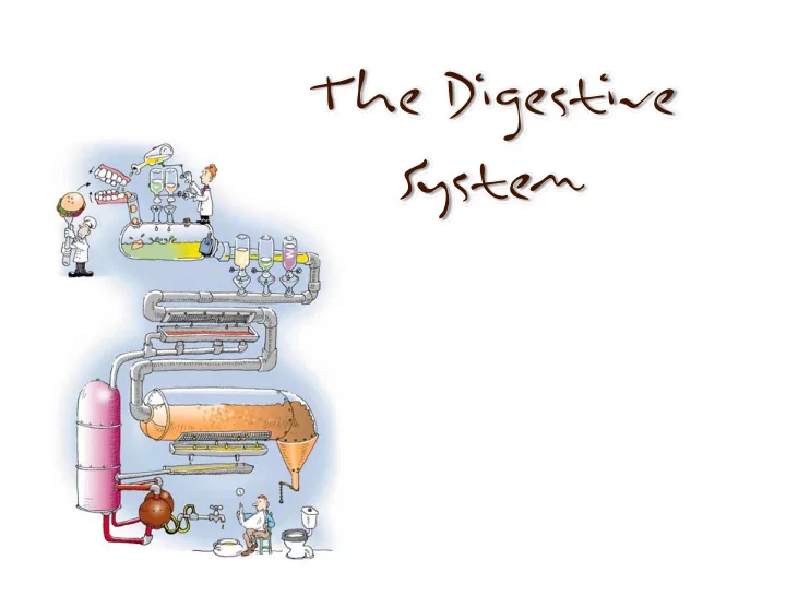
The Digestive System Overview of the Digestive System Organs are divided into two groups The alimentary canal Mouth, pharynx, and esophagus Stomach, small intestine, and large intestine (colon) Accessory digestive organs
groups – The alimentary canal
esophagus
intestine, and large intestine (colon)
– Teeth and tongue – Gallbladder, salivary glands, liver, and pancreas
Thin loose connective tissue, and epithelium which is continuous with the visceral peritoneum
Responsible for motility in GI tract Longitudinal and circular muscles (exceptions in stomach and colon). Contains nerves (myenteric plexus) for local control
Contains vessels, glands, nerves (submucosal plexus)
Composed of three layers:
tissue)
Peritoneum – a serous membrane
Visceral peritoneum – surrounds digestive
Parietal peritoneum – lines the body wall
Peritoneal cavity – the thin sandwiched space between the visceral and parietal peritoneum.
Figure 22.5a
layer of peritoneum
– Suspend & holds
– Sites of fat storage – Provides a route for circulatory vessels and nerves
Superficial view of the abdominal organs
Figure 22.6b
transverse colon reflected
– Greater omentum – Transverse mesocolon – Mesentery – Sigmoid mesocolon
Figure 22.6c
the abdominopelvic cavity
– Lesser omentum – Falciform ligament – Transverse mesocolon – Mesentery – Greater omentum
Figure 22.26d
– Behind the peritoneum
– Digestive organs that keep their mesentery
Figure 22.5b
– Mucosal layer composed of . . .
– Formed from orbicularis oris and buccinator muscles, respectively
– Salivary amylase & lingual lipase
Figure 22.8a
anchors the tongue
roof of the mouth
and uvula form the fauces (the arches that
Figure 22.8b
– Interlacing fascicles of skeletal muscle
– Motility ‐ grips food and repositions it – Communication ‐ helps form some consonants – Taste
the pharynx and larynx.
called papillae. There are three types of papillae:
– Filiform – Fungiform – Circumvallate
– First appear at 6 months of age
– Most erupt by the end of adolescence
– Way to indicate number and type of teeth. For Example: I 2/2, C 1/1, P 2/2, M 3/3), where I= incisors, C = canines, P = Premolars, M= Molar and the numbers represent the numbers of teeth in the upper and lower quadrant. To get the total number of teeth in a mouth, add all numbers and multiply by 2 = 32 teeth!
Figure 22.10
Deciduous (Baby) Teeth Permanent (Adult) Teeth
Figure 22.11
– Parotid, submandibular, and sublingual glands
Figure 22.12
for air and food
– Lined with stratified squamous epithelium – External muscle layer
constrictors – What about the nasopharynx?
– A 25 cm long simple muscular tube – Begins as a continuation of the pharynx at the upper esophageal sphincter – Travels through the posterior mediastinum – Joins the stomach inferior to the diaphragm – Ends at the lower esophageal sphincter (cardiac sphincter)
– Epithelium is stratified squamous epithelium – When empty
– Mucous glands
– Muscularis externa
– Most external layer – adventitia
Note the thick stratified squamous epithelium as well as the submucosoal and muscularis layers. Muscularis Submucosa Mucosa
(esophageal‐stomach junction)
– Due to churning action created by the additional
longitudinal and circular muscles)
amylase is denatured in acidic lumen
– Secretes pepsin – Functions under acidic conditions
– Water, alcohol, salts, aspirin
~ 1 qt. (slightly less than 1 liter)
inches long, by 6 inches wide
junction of esophagus
pyloric sphincter
Figure 22.14b
Microscopic Anatomy
Histology of Stomach