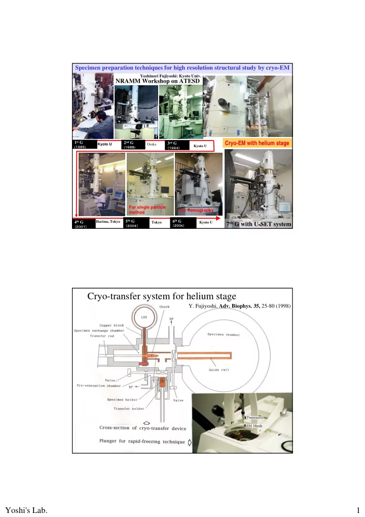

Specimen preparation techniques for high resolution structural study by cryo-EM Yoshinori Fujiyoshi: Kyoto Univ. NRAMM Workshop on ATESD 1 st G 2 nd G 3 rd G Cryo-EM with helium stage Kyoto U Osaka Kyoto U ( 1986 ) ( 1988 ) (1994) For single particle For Tomography method 5 th G 6 th G 4 th G 7 th G with U-SET system Harima, Tokyo Tokyo Kyoto U (200 6 ) (2004) (2001) Cryo-transfer system for helium stage Y. Fujiyoshi, Adv. Biophys. 35, 25-80 (1998) Yoshi's Lab. 1
Quick specimen exchange by our cryo-transfer system helps to optimize specimen preparation techniques Adv. Biophys. 35, 25-80 (1998) Structures of membrane proteins analyzed by cryo-EM AChR Gap Junction Bacteriorhodopsin Aquaporin-0 channel Nature, 389, 206-211 Nature, 423, 949- (1997) 955 (2003) Light-harvesting Nature, 438, 633-638 Aquaporin-1 PNAS, 104, 10034- complex (2005) 10039 (2007) MGST-1 Aquaporin-4 Nature, 407, 599- 605 (2000) Nature, 367, 614-621 JMB, 360, 934-945 Nature, 387 , 624- JMB, 355, 628-639 (2006) (1994) 627 (1997) (2006) Yoshi's Lab. 2
Requirements for structural study 1) Flat support Atomically flat carbon film Smooth Mo grid 2) Water evaporation (Dehydration, salt concentration) 3) Thinner embedding layer 4) Deformation by mechanical interaction 5) Suger embedding (Trehalose cushion) 6) Image deterioration by beam induced charge How could best EM specimens be prepared? Atomically flat carbon film Reason of the importance: blurring diffraction spots Yoshi's Lab. 3
Carbon film One spark with no spark Atomically smooth carbon film No spark Evaporaton on mica in high vacuum Carbon cluster Mo grid for minimizing cryo-crinkling Commercially avarable Mo grid Very smooth Mo grid Non circular Mo grid Yoshi's Lab. 4
Requirements for structural study 1) Flat support Atomically flat carbon film Smooth Mo grid 2) Water evaporation (Dehydration, salt concentration) 3) Thinner embedding layer 4) Deformation by mechanical interaction 5) Suger embedding (Trehalose cushion) 6) Image deterioration by beam induced charge How could best EM specimens be prepared? *AQP0: cataract, cell adhesion Significance of water channels *AQP1: fast water flow, many organs Involved in numerous physiological processes AQP2: trafficking according with V2R 13 water channels signal, cardiopathy AQP3 : glycerol, cure incision, beautification *AQP4: cell adhesion, array, manic- depressive AQP5: dry eye, salivation AQP6: permiate not water but anion AQP7: glycerol, a fat cell, obesity AQP8: glycerol, alimentary canal, pancreas, acinus, liver AQP9: glycerol, liver cell AQP10: glycerol, alimentary canal AQP11: NPA motif to PNC, nephrogenic diabetes insipidus Aquaporins in Human Body AQP12: NPAmotif to NPT Yoshi's Lab. 5
How is blood flow regulated without smooth muscle No vascular smooth muscle in brain Speaking English endfoot of by T. Nakata astrocyte Native speaker Glial lamellae of Hypothalamus Japanese M.A. Moghaddam, O.P. Ottersen, Nature Rev. Neurosci., 4, 991-1001(2003) Two dimensional crystals of AQP4 Expression by Sf9 cells & Purification A typical diffraction pattern from 2D-crystal ~70% Yet effective cryo-EM gave or or us its structure Double-layered 2D-crystals 45Å typical yield: ~3mg AQP4 from 1-liter of Sf9 cells Molecular arrangement in 2D-crystal Yoshi's Lab. 6
Diffraction pattern: 60 ˚ tilt Water evaporation causes deterioration of electron diffraction patterns: 60 ˚ tilt Dried crystal: untilt Water evaporation causes deterioration of electron diffraction patterns: untilt Yoshi's Lab. 7
Good crystal: untilt Dried crystal: untilt Water evaporation causes deterioration of electron diffraction patterns: untilt Yoshi's Lab. 8
Good crystal: untilt Size of Orthogonal arrays of AQP4 at endfeet of astrocyte by Neely J Orthogonal array D et al, Biochemistry (1999) 38: 11156-11163 The orthogonal array structure AQP4M23 AQP4M1 Yoshi's Lab. 9
Requirements for structural study 1) Flat support Atomically flat carbon film Smooth Mo grid 2) Water evaporation (Dehydration, salt concentration) 3) Thinner embedding layer 4) Deformation by mechanical interaction 5) Suger embedding (Trehalose cushion) 6) Image deterioration by beam induced charge How could best EM specimens be prepared? Reason why we need thinner embedding layer Mo grid Thick layer 2D-crystal Carbon film Mo grid Thin layer 2D-crystal Carbon film Yoshi's Lab. 10
Flow chart of electron crystallography Bended (undulated) crystal: 60 ˚ tilt Thicker layer makes crystals undulate and less clear diffraction spots in the direction perpendicular to the tilting axis Yoshi's Lab. 11
Good crystal: 60 ˚ tilt Thinner layer makes crystals less undulate and also gives better S/N ratio Requirements for structural study 1) Flat support Atomically flat carbon film Smooth Mo grid 2) Water evaporation (Dehydration, salt concentration) 3) Thinner embedding layer 4) Deformation by mechanical interaction 5) Suger embedding (Trehalose cushion) 6) Image deterioration by beam induced charge How could best EM specimens be prepared? Yoshi's Lab. 12
Structure of AQP4 Requirements for structural study 1) Flat support Atomically flat carbon film Smooth Mo grid 2) Water evaporation (Dehydration, salt concentration) 3) Thinner embedding layer 4) Deformation by mechanical interaction 5) Suger embedding (Trehalose cushion) 6) Image deterioration by beam induced charge How could best EM specimens be prepared? Yoshi's Lab. 13
Effect of Trehalose cushion Low trehalose cushion Higth trehalose cushion Trehalose embedding method Yoshi's Lab. 14
Gap Junction channel: Cx26 GJ channels permiate peptides of 1.8kD, Electrical Chemical gating mechanism for blocking ions ? synapse synapse Long standing 20 Å questions Multiple gating mechanisms by voltage, calcium ion, phosphorylation, pH 2D-crystal of Gap Junction channels Surprizingly 3- membrane layers! Crytoplasmic structures at Mem-1 and -3 are easily deformed but these at Mem-2 are protected ! Yoshi's Lab. 15
Plug density in Cx26 channel ! 栓:Plug Plug PNAS, 104 , 10034-10039 (2007) A New density 栓:Plug Stereoscopic view at cytoplasmic side B-loop B-loop B-loop B-loop Structure at cytoplasmic side is related with caracteristic feature of each Cx: White arrows show B loop between Helices2-3 green arrows indicate N-terminal loops interacting with the B loops Yoshi's Lab. 16
Single particle analysis Na-channel, Nature, 409, 1047-1051 (2001) IP 3 R ., J. Mol Biol., 336, 155-164 (2004). Vestibules Vestibules Single particle analysis of TRPC3 Neuronal differentiation, blood vessel constriction & immune cell maturation Yoshi's Lab. 17
Neuro-muscular junction ACh Nature, 423, 949-955 (2003) Ach esterase AChR ← Vestibules Na + Na + -channel ← (B.Hille, 2 nd Ed.,SinauerAssociates, Sunderland, MA, 1992) IIIIIIIIIIIIIIIIIIIIIIIIIIIIIIIIIIIIIIIIIIIIIIIIIIIIIIIII Ach: Positively charged IIIIIIIIIIIIIIIIIIIIIIIIIIIIIIIIIIIIIIIIIIIIIIIIIIIIIIIII Negative charge:Ion filter Negative charge Ion Rapsyn filter Rapsyn Image of a tubular crystal embedded in vitreous ice Amorphous ice Tubular crystal S c a n a r e a Holey carbon support film 1000Å No interaction which induces deformation Yoshi's Lab. 18
2D-crystal of Gap Junction channels Molecules at outer 1- & 3-membrane layers are deformed but minimized by trehalose cushion Crytoplasmic structures at Mem-1 and -3 are easily deformed but these at Mem-2 are protected by ! Requirements for structural study 1) Flat support Atomically flat carbon film Smooth Mo grid 2) Water evaporation (Dehydration, salt concentration) 3) Thinner embedding layer 4) Deformation by mechanical interaction 5) Suger embedding (Trehalose cushion) 6) Image deterioration by beam induced charge How could best EM specimens be prepared? Yoshi's Lab. 19
Image of a tubular crystal embedded in vitreous ice Amorphous ice Tubular crystal S c a n a r e a Holey carbon support film 1000Å N. Unwin’s image Gold particles embedded in ice Yoshi's Lab. 20
Thermal conductivity Gold particles embedded in ice Visible area Solid N 2 Bubble Gold particle Vitreous ice Gold Bubble Carbon film Mo grid Yoshi's Lab. 21
Gold particles embedded in ice Pre-irradiation technique Beam induced movement of a particle embedded in ice Yoshi's Lab. 22
Structure analysis from tubular crystals Image of tubular crystal 3D-structure analysis 1.unbending 2.averaging Nature, 423, 949-955 (2003) Requirements for structural study 1) Flat support Atomically flat carbon film Smooth Mo grid 2) Water evaporation (Dehydration, salt concentration) 3) Thinner embedding layer 4) Deformation by mechanical interaction 5) Suger embedding (Trehalose cushion) 6) Image deterioration by beam induced charge Image shift How could best EM specimens be prepared? Yoshi's Lab. 23
3 Å Structure of bR Nature, 389, 206-211 (1997) J. Mol. Biol., 286, 861-882 (1999) Very difficult to take good Image shift caused by charge up images at tilted conditions Yoshi's Lab. 24
Recommend
More recommend