
SLIDE 1
The Basis of Evidence: Transfer and Persistence Developed by Edmund - - PowerPoint PPT Presentation
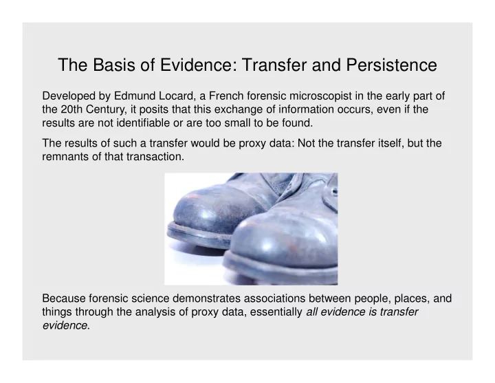
The Basis of Evidence: Transfer and Persistence Developed by Edmund Locard, a French forensic microscopist in the early part of the 20th Century, it posits that this exchange of information occurs, even if the results are not identifiable or are
absent
abundant fusi
cut broken split
coarse
thickness
protrusion
cells prominent
size of granules
bleached
cracked cuticle
Microscopic Mitochondrial Association 80 97 Inconclusive 37 3 Exclusion 19 64 No Exam 34 6
#
%$ % !
Results of Microscopic and Mitochondrial DNA Analyses by Method
casework hairs was carried out.
– With increasing age of the hair, the likelihood of obtaining a full profile decreased – With increasing color and diameter of the hair, the likelihood of obtaining a profile increased. – Full or partial profiles were obtained on more than 80% of 114 hairs 1.0 cm. Mixtures were observed in 8.7% of hairs tested.
exterior surface contamination that could not be sufficiently cleaned prior to extraction, since the overall level of laboratory contamination was low.
sites were observed.
1Melton, et al., JFS V50, N1, 2005 2Roberts and Calloway, JFS V52, N1, 2007