
SLIDE 1
Sickle Cell and the Eye
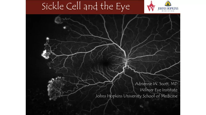
Adrienne W. Scott, MD Wilmer Eye Institute Johns Hopkins University School of Medicine
Sickle Cell and the Eye Adrienne W. Scott, MD Wilmer Eye Institute - - PowerPoint PPT Presentation
Sickle Cell and the Eye Adrienne W. Scott, MD Wilmer Eye Institute Johns Hopkins University School of Medicine Disclosures Allergan, Inc. (Consultant) No financial interest in this subject matter Sickle Cell and the Eye- Take Home
Adrienne W. Scott, MD Wilmer Eye Institute Johns Hopkins University School of Medicine
Schundeln et.al, Journal of Pediatrics, 2014.
Top photo by Andrew R. Marks, M.D.
Stage IV: vitreous hemorrhage
Stage IV: vitreous hemorrhage Seafans visible despite vitreous heme
Stage V: vitreous hemorrhage Combined TRD/RRD
Image Courtesy of the American Society of Retina Specialists (ASRS) image Bank
Stage 3 Stage 4 Stage 5
Pre-treatment Pre-treatment Post-treatment Post-treatment
– Refractive error separate
– Not sure, a thorough dilated exam will pick up serious retinal pathology
– Probably, and will increase as they age
– Probably not, risk is low, but subtle vision loss may be underdiagnosed
– At least annually, age 5 and older, NHLBI consensus suggests age 10 – Floaters, or vision loss, HbSC/Beta thal