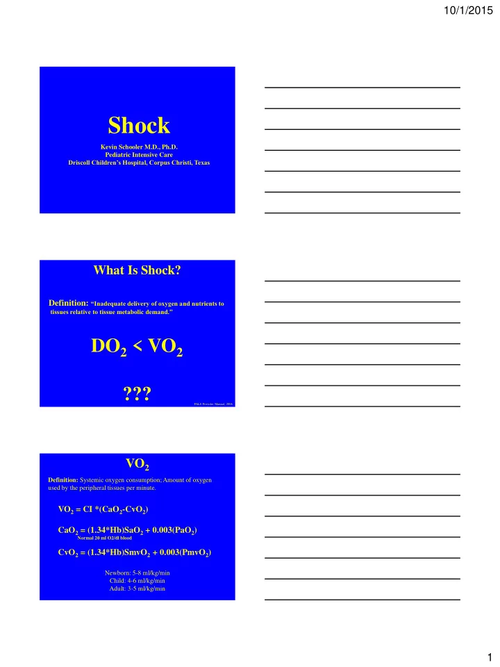
Shock Kevin Schooler M.D., Ph.D. Pediatric Intensive Care Driscoll - - PDF document

shock
SMART_READER_LITE
LIVE PREVIEW

Shock Kevin Schooler M.D., Ph.D. Pediatric Intensive Care Driscoll - - PDF document

10/1/2015 Shock Kevin Schooler M.D., Ph.D. Pediatric Intensive Care Driscoll Childrens Hospital, Corpus Christi, Texas What Is Shock? Definition: Inadequate delivery of oxygen and nutrients to tissues relative to tissue metabolic


slide-1
SLIDE 1

10/1/2015 1

Shock

Kevin Schooler M.D., Ph.D. Pediatric Intensive Care Driscoll Children’s Hospital, Corpus Christi, Texas

What Is Shock?

Definition: “Inadequate delivery of oxygen and nutrients to

tissues relative to tissue metabolic demand.”

PALS Provider Manual, 2006

DO2 < VO2 ???

VO2

VO2 = CI *(CaO2-CvO2) CaO2 = (1.34*Hb)SaO2 + 0.003(PaO2)

Normal 20 ml O2/dl blood

CvO2 = (1.34*Hb)SmvO2 + 0.003(PmvO2)

Definition: Systemic oxygen consumption; Amount of oxygen used by the peripheral tissues per minute. Newborn: 5-8 ml/kg/min Child: 4-6 ml/kg/min Adult: 3-5 ml/kg/min

slide-2
SLIDE 2

10/1/2015 2

DO2

DO2 = CI*(CaO2) CaO2 = (1.34*Hb)SaO2 + 0.003(PaO2)

Normal 20 ml O2/dl blood

DO2 = CI *[(1.34*Hb)SaO2 + 0.003(PaO2)]

Definition: Systemic oxygen delivery; Amount of oxygen delivered to the peripheral per minute. Normal: 5 times VO2 regardless of size

BP is NOT a part of the DO2 equation!!! Notice: Relationship Between DO2 and VO2

Adapted from R.H. Bartlett, “Physiology of Extracorporeal Life Support.”, ECMO Support in Critical Care, 2 nd Edition, 2000

VO2 (ml/kg/min) DO2 (ml/kg/min) 10 20 15 5 5 4 3 2 1 A B C D

Increase in DO2 (A-B) or decrease in DO2 (A-C), does not affect VO2 unless DO2:VO2 drops below 2:1 (A-D)

slide-3
SLIDE 3

10/1/2015 3

Translation: If tissues use (VO2)

  • xygen faster than it is delivered

(DO2) the patient is in shock.

DO2 < VO2

By More Than 2:1

So… Now What?

Glucose G6P Pyruvate Lactate NAD + ATP

Glucose 2 Lactate + 2 ATP + 2 NAD

Anerobic Glycolysis

Aerobic Glycolysis

Glucose 2 Pyruvate + 2 ATP + 2 NADH

The Core Metabolic Pathway

slide-4
SLIDE 4

10/1/2015 4

  • Great. But how does

that help me recognize and treat shock?

Stages of Shock

Compensated- Blood Pressure Maintained

  • end organ function not yet showing evidence of

being terminally compromised

Uncompensated- Blood pressure falls

  • end organs dysfunction becomes clinically significant

Irreversible- Multi-organ failure

  • extensive damage that can no longer be repaired

What Is Normal SBP?

Newborn: SBP >= 60 1 month – 1 year: SBP >= 70 > 1 year SBP >= 70 + 2 * Age in years > 10 years SBP >= 90

Adapted from PALS Provider Manual, 2002

slide-5
SLIDE 5

10/1/2015 5

What About MAP?

5th percentile = (1.5 * years) + 40 50th percentile = (1.5 * years) + 55

I thought BP was NOT a part

  • f the

definition of shock!!! But... Why Is Low BP A Late Sign Of Shock?

Since Q = DP/SVR Then MAP a CO * SVR

slide-6
SLIDE 6

10/1/2015 6

SVR MAP CO Normal 10% 20% 30% Blood V

  • lume Lost

Compensated vs. Uncompensated Shock?

Adapted from PALS Provider Manual, 2002

So Let’s Change That: Stages of Shock

Reversible- end organ function not yet showing evidence of

being terminally compromised

Irreversible- Multi-organ failure

  • extensive damage that can no longer be repaired
  • BP is lost at some point

What End Organs Can We Evaluate Easily?

Brain: Mental status normal? Changing? Declining? Kidneys: Urine becoming concentrated?

UOP volume falling off?

Skin: Warm? Cold? Capillary refill?

  • reflects CO

Heart: Tachycardia worsening? Improving?

  • reflects volume status or dysfunction

Lungs: Tachypnea worsening? Improving?

  • reflects acidosis
slide-7
SLIDE 7

10/1/2015 7

“Prevent end-organ injury and halt progression to cardiopulmonary failure and cardiac arrest.”

Therefore: Treatment Goals

PALS Provider Manual, 2006

Lowering VO2

Sedate: decreases pain, agitation and neurologic electrical activity Intubate: eliminating WOB, decreases CO by 20-30% Decrease Temperature: higher T is higher metabolic demand Paralyze: eliminates muscle activity

What Are The DO2 Components?

CO = HR * SV

Preload Contractility Afterload Rhythm

DO2 = CI *[(1.34*Hb)SaO2 + 0.003(PaO2)] CI = CO/BSA

Hypoxia Anemia

slide-8
SLIDE 8

10/1/2015 8

Relationship Between Hb, PO2 And O2 Content

Adapted from R.H. Bartlett, “Physiology of Extracorporeal Life Support.”, ECMO Support in Critical Care, 2 nd Edition, 2000

O2 Content (ml/dl) PO2 25 20 15 10 5 50 100 75 25 125 150 Hb 15 Hb 10 Hb 7.5 V A

In Other Words:

Doubling Hgb Will Double The DO2!

Shock Reversal Requires You To Know The Type Of Shock You Are Dealing With!

slide-9
SLIDE 9

10/1/2015 9

Shock Classification

Hypovolemic Cardiogenic Distributive Obstructive Dissociative

O2 O2 O2 O2 O2 O2 O2 O2 O2 O2 O2 O2 O2 O2 O2 O2 O2 O2 O2 O2

DO2 VO2

Balanced Input and Output

Mechanisms of BP Homeostasis

1) Hyperacute:

  • Carotid and aortic baroreceptors
  • sense fall in blood pressure
  • Medullary stimulus increases Sympathetic (adrenergic) and decreases

Parasympathetic (vagal) tone

  • HR and SVR increase to maintain BP
  • Acidosis sensed in carotid bodies
  • HR and SVR increases to improve BP
  • RR increases to alleviate acidosis
  • Serum Osmolality rises
  • ADH released from pituitary
  • SVR increases
  • water channels in collecting ducts close to increase volume

Blood Pressure Falls

slide-10
SLIDE 10

10/1/2015 10

2) Acute:

  • Decreased blood pressure causes decreased perfusion of kidney JG apparatus
  • Renin secretion increases
  • Renin cleaves Angiotensinogen to Angiotensin I
  • ACE cleaves Angiotensin I to Angiotensin II
  • Angiotensin II
  • Direct increase in SVR
  • Pituitary gland secretes ADH
  • SVR increases
  • water channels in collecting ducts close to increase

volume

  • aldosterone secreted from adrenal cortex
  • sodium retained to increase volume

Mechanisms of BP Homeostasis

3) Acute:

Increased water volume increases RAP

  • CO increases
  • Increased water volume increases EDV
  • CO increases

Continued Stress

Increased EDP Pulmonary Edema Increased Ventilator Support Decreased Venous Return Decreased CO More volume needed etc…. O2 O2 O2 O2

O2 O2 O2 O2 O2

O2 O2 O2 O2 O2 O2 O2 O2

O2

O2

DO2

VO2

O2 O2 O2 O2 O2 O2 O2 O2 O2 O2 O2

Hypovolemic Shock

Pipes are empty

slide-11
SLIDE 11

10/1/2015 11

CO = HR * SV

Preload Contractility Afterload Rhythm

DO2 = CI *[(1.34*Hb)SaO2 + 0.003(PaO2)] CI = CO/BSA

Hypoxia Anemia

Hypovolemic Shock Hypovolemic Shock

Etiology: Decrease in total circulating volume Subtypes:

Hemorrhagic Shock Burns Osmotic Diuresis (DKA) Third Spacing Diarrhea/Vomiting

Diagnosis: ↑ Rate ↓ Preload

↑ ↔ Contractility, ↑ Afterload

Treatment: Blood, Fluid Symptoms: tachycardia, tachypnea, cold extremities,

narrow pulse pressure, prolonged CR

O2 O2 O2 O2 O2 O2 O2 O2 O2 O2 O2 O2 O2 O2 O2 O2 O2 O2 O2 O2

Cardiogenic Shock

Pump is Broken

DO2

VO2

slide-12
SLIDE 12

10/1/2015 12

CO = HR * SV

Preload Contractility Afterload Rhythm

DO2 = CI *[(1.34*Hb)SaO2 + 0.003(PaO2)] CI = CO/BSA

Hypoxia Anemia

Cardiogenic Shock Cardiogenic Shock

Etiology: Inadequate cardiac output Diagnosis: ↓ or ↑ Rate

↔ Preload ↓ Contractility ↑ Afterload

Treatment: Optimization of preload, afterload,

contractility, rhythm, and rate

Symptoms: tachycardia, tachypnea, cold extremities,

narrow pulse pressure, prolonged CR, weak pulses diaphoresis, CHF symptoms, cyanosis

O2 O2 O2 O2 O2 O2 O2 O2 O2 O2 O2

O2 O2 O2 O2 O2 O2 O2 O2 O2

Distributive Shock

Pipes Are Too Big

slide-13
SLIDE 13

10/1/2015 13

CO = HR * SV

Preload Contractility Afterload Rhythm

DO2 = CI *[(1.34*Hb)SaO2 + 0.003(PaO2)] CI = CO/BSA

Hypoxia Anemia

Distributive Shock Distributive Shock

Etiology: Inappropriate distribution of blood volume Subtypes:

Septic shock Anaphylactic shock Neurogenic shock

Diagnosis: ↑ ↔ Rate ↓ ↔ Preload,

↓ ↔ Contractility, ↔ ↓ Afterload

Treatment: Fluid, vasopressor etc. Symptoms: tachycardia, tachypnea, warm or cold extremities,

wide or narrow pulse pressure, brisk or prolonged CR, weak or strong pulses

Dellinger et al. “Surviving Sepsis Campaign: International Guidelines for Management of Severe Sepsis and Septic Shock: 2012.” Crit .Care Med . V41: 2, 2013

Septic Shock

slide-14
SLIDE 14

10/1/2015 14

Relationship Between DO2 and SVO2

Adapted from R.H. Bartlett, “Physiology of Extracorporeal Life Support.”, ECMO Support in Critical Care, 2 nd Edition, 2000

DO2 (ml) 600 900 300 1200 VO2 (ml) 240 180 120 60 300 2 50 3 66 4 75 5 80 DO2/VO2 ratio: SVO2:

Anaphylactic Shock

Epinephrine: 0.15 mg IM < 30 Kg

0.3 mg IM >30 Kg

Albuterol:

0.5 mg/kg/hr

Antihistamine:

H1-diphenhydramine H2-ranitidine

Corticosteroids: Solumedrol 1 mg/kg

Neurogenic Shock

Volume & Norepinephrine

slide-15
SLIDE 15

10/1/2015 15

Obstructive Shock

O2 O2 O2 O2

VO2

Pipes Are Clogged

DO2

CO = HR * SV

Preload Contractility Afterload Rhythm

DO2 = CI *[(1.34*Hb)SaO2 + 0.003(PaO2)] CI = CO/BSA

Hypoxia Anemia

Obstructive Shock Obstructive Shock

Etiology: Impaired CO caused by physical obstruction

  • f blood flow

Subtypes:

Cardiac Tamponade Tension Pneumothorax Ductal Dependent CHD Pulmonary embolism

Treatment: Fluid and Relief of Obstruction Symptoms: tachycardia, tachypnea, cold extremities,

narrow pulse pressure, prolonged CR, weak pulses

slide-16
SLIDE 16

10/1/2015 16

Relieve The Obstrtuction

Cardiac Tamponade- emergent pericardiocentesis Tension Pneumothorax- 18-20G needle decompression,

  • ver third rib mid-clavicular line

Ductal Dependent CHD- Prostaglandin E1 Pulmonary Embolus- Anticoagulants, thrombolytics

Dissociative Shock

Etiology: Impaired oxygen delivery despite normal heart,

blood flow, blood volume and vascular tone

Subtypes:

Poisons (Cyanide, CO) Inborn Errors of Metabolism Abnormal hemoglobin

Treatment: Reverse underlying cause Symptoms: tachycardia, tachypnea, acidosis