
SLIDE 1
BONE & JOINT INFECTIONS
Henry F. Chambers, MD
SEPTIC ARTHRITIS Native Joint
Curr Rheumatol Rep 15:332, 2013; Best Prac Res Clin Rheumatol 25:407, 2011
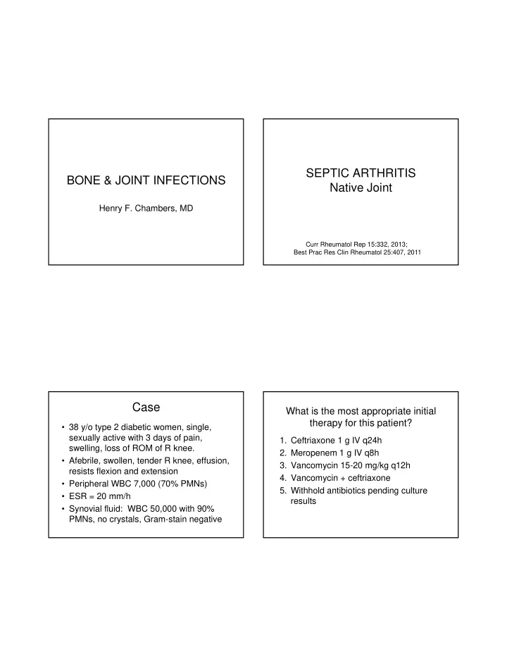
Case
- 38 y/o type 2 diabetic women, single,
sexually active with 3 days of pain, swelling, loss of ROM of R knee.
- Afebrile, swollen, tender R knee, effusion,
resists flexion and extension
- Peripheral WBC 7,000 (70% PMNs)
- ESR = 20 mm/h
- Synovial fluid: WBC 50,000 with 90%
PMNs, no crystals, Gram-stain negative
What is the most appropriate initial therapy for this patient?
- 1. Ceftriaxone 1 g IV q24h
- 2. Meropenem 1 g IV q8h
- 3. Vancomycin 15-20 mg/kg q12h
- 4. Vancomycin + ceftriaxone
- 5. Withhold antibiotics pending culture