
SLIDE 1
New Therapies in Rheumatoid Arthritis 1
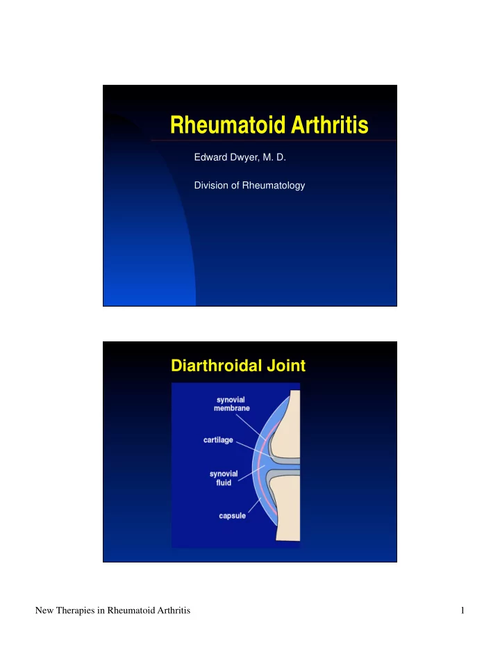
Rheumatoid Arthritis
Edward Dwyer, M. D. Division of Rheumatology
Rheumatoid Arthritis Edward Dwyer, M. D. Division of Rheumatology - - PDF document
Rheumatoid Arthritis Edward Dwyer, M. D. Division of Rheumatology Diarthroidal Joint New Therapies in Rheumatoid Arthritis 1 Diarthroidal Joint in Rheumatoid Arthritis Normal Synovium New Therapies in Rheumatoid Arthritis 2 Synovium in
New Therapies in Rheumatoid Arthritis 1
Edward Dwyer, M. D. Division of Rheumatology
New Therapies in Rheumatoid Arthritis 2
New Therapies in Rheumatoid Arthritis 3
New Therapies in Rheumatoid Arthritis 4
Pannus composed of macrophages and mesenchymal cells which erode into cartilage and bone
T cells
CD4 TH1 phenotype (IFN-, IL-2)
Macrophages
TNF and IL-1
B cells
Rheumatoid Factor Anti-Cyclic Citrullinated Peptide Ab (anti-CCP Ab)
New Therapies in Rheumatoid Arthritis 5
Cytokine Produced by Activity
IL-1 IL-6 IL-15 IL-17 IL-18 IL-23 IL-32 M M, Ly, Fibr M, Syn, Endo TH17 cells M M M, Ly “TLR-like”; activates NF-B Induces IL-17; stimulates bone resorption “IL-2-like”; stimulates TH1 polarization Induces TNF-, IL-1, RANKL “TLR-like”; activates NF-B IL-12 family member; induces IL-17 Induces TNF, IL-1, IL-6, and chemokines
Prevalence of 1% in most populations Age of onset: 30-50 yrs Sex: F:M 3:1
New Therapies in Rheumatoid Arthritis 6
Sex
F:M 3:1
Family History:
Monozygotic twins: RR = 8
Concordance rate: 30%
Dizygotic twins: RR = 2-3.4 First degree relative: RR = 1.5
MHC association accounts for 40% genetic risk
Alleles of the DR1 locus are responsible for
increased risk to RA
Alleles of DR1 chain that confer increased risk
exhibit a “shared epitope” of amino acid sequence in the the third hypervariable region from amino acids 70-74
e.g., DR1*0401, DR1*0404, DR1*0101
In some populations >95% of patients with RA
exhibit this “shared epitope”
New Therapies in Rheumatoid Arthritis 7
Third Hypervariable Region
“Shared Epitope” Third Hypervariable Region Sequence:
70 71 72 73 74
IgM antibody with specificity for the Fc region of IgG
New Therapies in Rheumatoid Arthritis 8
Rheumatic Diseases
SLE, Sjogren’s syndrome
Viral Infections
HCV, HIV
Bacterial Infections
SBE, TB, syphilis, leprosy
Neoplasms
Lymphoproliferative diseases
Present in 3% general population
Sensitivity: 70% Specificity: 60%
New Therapies in Rheumatoid Arthritis 9
Arginine Citrulline
Post-translational modification of arginine as a consequence of cell death and inflammation, i.e., oxidative stress Proteins derived from synovial tissue in RA
exhibit enhanced citrullination
Patients with RA have high titers of
autoantibodies directed against proteins with citrulline residues
e.g., anti-CCP Assay (ELISA assay)
New Therapies in Rheumatoid Arthritis 10
Sensitivity: 70% Specificity: 95%
anti-CCP
New Therapies in Rheumatoid Arthritis 11
Morning stiffness (> 1 hour) Arthritis of 3 or more joint areas (polyarticular) Arthritis of hand joints Symmetric arthritis Rheumatoid nodules Rheumatoid Factor in serum Radiographic changes:
Periarticular demineralization of bone (early) Marginal erosions (later)
*1987 American College of Rheumatology Revised Criteria for the Classification of RA
4 of 7 criteria should be present to diagnose Rheumatoid Arthritis
New Therapies in Rheumatoid Arthritis 12 Polyarticular Arthritis of hand joints most common Metacarpophalangeal joints (MCPs) Proximal interphalangeal joints (PIPs) Never Distal interphalangeal joints (DIPs) Symmetric arthritis
Less commonly involves: Toes, wrists, knees Least commonly involves: Shoulders, hips
New Therapies in Rheumatoid Arthritis 13
New Therapies in Rheumatoid Arthritis 14
New Therapies in Rheumatoid Arthritis 15
New Therapies in Rheumatoid Arthritis 16
Early changes
No abnormalities
Initial changes
Periarticular osteopenia secondary to cytokine-
induced bone loss
Later changes
Marginal erosions at periphery of joint
(cartilage-pannus interface)
Advanced changes
Joint space narrowing, subluxation
Radiographic Progression of MCP Joint Destruction
New Therapies in Rheumatoid Arthritis 17
New Therapies in Rheumatoid Arthritis 18
Extra-articular manifestations of RA are
generally found in those patients who have relatively severe articular disease
Extra-articular disease is associated
with increased morbidity and mortality
New Therapies in Rheumatoid Arthritis 19
Necrotic core Palisading rim of tissue macrophages and T cells
New Therapies in Rheumatoid Arthritis 20
New Therapies in Rheumatoid Arthritis 21
Interstitial infiltration of macrophages and T cells resulting in pulmonary fibrosis
New Therapies in Rheumatoid Arthritis 22
Rheumatoid Arthritis Neutropenia Splenomegaly
1-2% Rheumatoid Arthritis patients 1/3 have expansion of CD3+CD8+ Large
Granular Lymphocytes in peripheral smear
Increased risk for infections and non-
Hodgkins lymphoma
New Therapies in Rheumatoid Arthritis 23
Reduce or eliminate pain Prevent or retard joint destruction Maintain musculoskeletal functional status Prevent or retard development of extra-
articular manifestations of disease
New Therapies in Rheumatoid Arthritis 24
Joint-space narrowing and erosion are seen in
67% of patients within the first 2 yrs of disease
Joint-space narrowing and erosion are seen in
77% of patients within the first 5 yrs of disease
Progression is most rapid during the first 5 yrs
“The majority of patients with newly diagnosed RA should be started on Disease-Modifying Anti-Rheumatic Drug (DMARD) therapy within 3 months of diagnosis.”
Arthritis & Rheumatism, 46(2), 328-46, 2002
New Therapies in Rheumatoid Arthritis 25
Prostaglandin inhibitors that exhibit
analgesic and anti-inflammatory effects
e.g., aspirin, ibuprofen, naproxen
NSAIDS do not inhibit or retard the
progression of articular destruction in Rheumatoid Arthritis
Useful for symptom management only
Methotrexate: Folic acid analog
that inhibits dihydrofolate reductase, an enzyme active in nucleic acid synthesis
New Therapies in Rheumatoid Arthritis 26
synthesis and therefore the proliferation of immune cells that mediate inflammation.
results in increased production of adenosine which mediates immunosuppressive and anti-inflammatory effects.
New Therapies in Rheumatoid Arthritis 27
Definitely improves symptoms and function,
and retards joint destruction in a significant percentage of patients.
However, < 50% of patients experience a
sustained remission on methotrexate alone
Anticytokine agents
Anti-TNF agents
Etanercept (Enbrel) Infliximab (Remicade) Adalimumab (Humira)
Anti-IL 1
Anakinra (Kineret)
B cell depleting agent
Anti-CD20
Rituximab (Rituxan)
Costimulatory inhibitor
Anti-B7 (CD80)
Abatacept (Orencia)
New Therapies in Rheumatoid Arthritis 28 TNF-
that is composed of three identical subunits
macrophages
cytes, and osteoclasts
pro-inflammatory cytokines, (e.g., IL-1 and IL-6) and matrix metalloproteinases
Etanercept
New Therapies in Rheumatoid Arthritis 29
activation
etanercept allows it to be 1000% times more efficient than the monomeric structure in neutralizing TNF
markedly prolongs the half-life
Etanercept
Subcutaneous Injection:
50 mg q. week
Half-life of 4 days Generally administered in addition to
methotrexate
New Therapies in Rheumatoid Arthritis 30
Intravenous Infusion of 3 mg/kg every 8
weeks
Development of anti-chimeric antibodies
to the murine region of the molecule is partially inhibited by the maintenance of methotrexate therapy
New Therapies in Rheumatoid Arthritis 31
IgG1 fully “humanized” monoclonal antibody
generated through application of phage display library technology
Avoids generation of anti-chimeric antibodies
Subcutaneous Injection:
40 mg q. 2 wks
Half-life: 2 weeks In addition to methotrexate
maintenance therapy
New Therapies in Rheumatoid Arthritis 32
Rapid onset of action (1-2 weeks) Sustained clinical response Retards (arrests?/reverses?) joint
destruction
Well tolerated
Reactivation of Latent Tuberculosis
TNF is an important cytokine in the immune
response to Mycobacterium tuberculosis
All patients need to be screened for previous
exposure to M. tuberculosis before initiating therapy with any anti-TNF agent
Those that exhibit a positive response to
PPD (purified protein derivative) need to be treated with antituberculous therapy
New Therapies in Rheumatoid Arthritis 33
IL 1 receptor antagonist (IL-1 Ra)
Naturally occurring protein produced by
macrophages at sites of inflammation that inhibits IL-1 induced activation
Anakinra (Kineret)
Human recombinant form of IL-1 Ra produced
in vitro
Subcutaneous injection
100 mg per day
Half-life: 6 hours Very modest efficacy
New Therapies in Rheumatoid Arthritis 34
monoclonal antibody targeting CD20 expressed on B cells
specific cell surface molecule expressed from pre-B cells to mature B cells (not expressed on plasma cells)
Mechanism of action in RA?
Does not interfere with autoantibody
production (e.g., RF or anti-CCP Ab) since it does not target plasma cells
Hypothesis: Rituximab reduces the role of B
cells that function as antigen presenting cells in presenting self-peptides to T cells in RA
New Therapies in Rheumatoid Arthritis 35
Intravenous infusion of 1000 mg
every 6 months
Half-life: 2-3 weeks B cell depletion lasts 4-6 months
New Therapies in Rheumatoid Arthritis 36
Extracellular CTLA-4 + IgG1 Constant Region
New Therapies in Rheumatoid Arthritis 37
Abatacept
CD28 CTLA4 exhibits 50-fold increased affinity for B7 vs. CD28 B7
Administration: Intravenous infusion of 10
mg/kg per month
Half-life: 15 days
New Therapies in Rheumatoid Arthritis 38
Cytokine Produced by Activity
IL-1 IL-6 IL-15 IL-17 IL-18 IL-23 IL-32 M M, Ly, Fibr M, Syn, Endo TH17 cells M M M, Ly “TLR-like”; activates NF-B Induces IL-17; stimulates bone resorption “IL-2-like”; stimulates TH1 polarization Induces TNF-, IL-1, RANKL “TLR-like”; activates NF-B IL-12 family member; induces IL-17 Induces TNF, IL-1, IL-6, and chemokines
Edward Dwyer, M. D. Division of Rheumatology