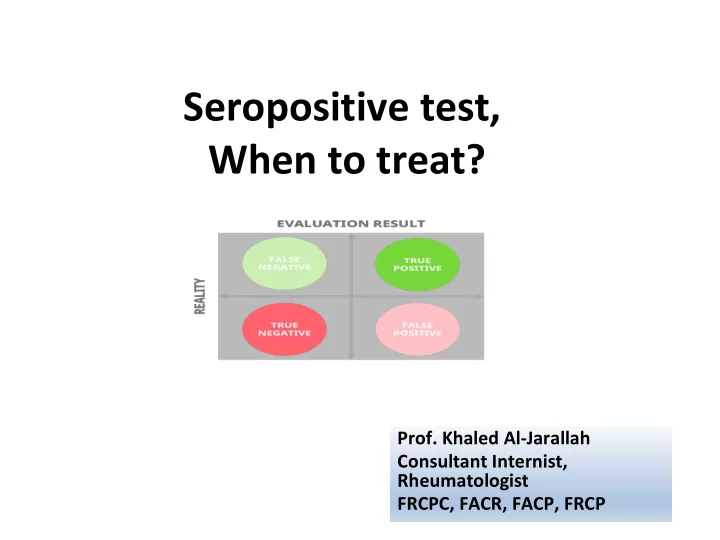
SLIDE 1 Seropositive test, When to treat?
Consultant Internist, Rheumatologist FRCPC, FACR, FACP, FRCP
SLIDE 2
Disclosures
I have no conflict of interest to declare
SLIDE 3 References
- Clinical Reasoning Series , Rahul patwari, 2017.
- Medow MA, Lucey CR.BMJ Evidence-Based Medicine. A
qualitative approach to Bayes theorem. 2011;16:163-167.
- Schur Peter, Laboratory testing for Diagnosis, Management
- f Patients with Rheumatic Disease. The Rheumatologist,
Dec 1,2014.
SLIDE 4 Referral Scenarios
- 48-year-old female referred from polyclinic
with 2 weeks history of generalized body ache & +RF 1:80 R/O Rheumatoid arthritis?
- Asymptomatic 25-year-old female, referred from
polyclinic after employment medical check-up with lab tests showing ANA 1: 640
SLIDE 5
Lab test
❑ For informing us of an emerging disease ❑ Diagnose a specific disease ❑ Predict prognosis ❑ Biomarker of disease ❑ Monitor of disease activity
SLIDE 6
!!
Why was the test requested?
SLIDE 7
Good referral
❑ 34-year-old female ❑ 12 week history of fatigue, joints pain&swelling in both hands.
SLIDE 8
SLIDE 9 Sensitivity, Specificity
Autoantibody positivity alone does not make a diagnosis Similarly, antibody negativity does not exclude a diagnosis ❑If highly sensitive, diagnosis can be excluded in case of negativity ❑If test is highly specific, diagnosis confirmed in case of positivity
SLIDE 10
SLIDE 11
SLIDE 12 Pre and Post test probability
Birtane M. et al 2017
The likelihood nomogram used in SLE with an antinuclear antibody test
SLIDE 13
SLIDE 14 Categorical probabilities A qualitative approach to Bayes theorem
Categorical probability Numerical probability Very unlikely Less likely than 10% Unlikely Between 10% and 33% Uncertain Between 34% and 66% Likely Between 67% and 90% Very likely More likely than 90%
Catherine R Lucey, Medow MA, Lucey CR.BMJ Evidence-Based Medicine 2011;16:163-167
SLIDE 15
SLIDE 16 Clinical reasoning approach
- Construct patient script (data gathering)
- Construct disease script (disease population
studies)
- Match= treat
- Mismatch= rule-out
- Uncertain= test
SLIDE 17 Clinical Reasoning , Rahul patwari, 2017
Clinical reasoning approach
SLIDE 18 Catherine R Lucey, Medow MA, Lucey CR.BMJ Evidence-Based Medicine 2011;16:163-167
SLIDE 19 Clinical Reasoning , Rahul patwari, 2017
SLIDE 20 Clinical Reasoning , Rahul patwari, 2017
SLIDE 21 Clinical Reasoning , Rahul patwari, 2017
SLIDE 22 Lupus in Arab world - disease script
Adwan M, Arch Rheumatol 2018;33(4);455-463
SLIDE 23 ANA ds DNA SSA /Ro aCL total Ribonucleoprotein Lupus Anti Smith SSB/La RF
Autoantibody Percentage
Frequency of autoantibodies in Arab patients with systemic lupus erythematosus
Adwan M, Arch Rheumatol 2018;33(4);455-463
Lupus in Arab world – disease script
SLIDE 24 Frequency of systemic lupus erythematosus manifestations in Arab world.
Percentage
Arthralgia/arthritis Anemia Fatigue Malar rash Renal Hemolytic anemia Photosensitivity Fever Alopecia Leucopenia Serositis Oral ulcers Neuropsychiatric Thrombocytopenia Lymphadenopathy Pulmonary Cardiac APS Discoid rash Raynaud’s phenomenon Vasculitis Thrombosis Myositis
Manifestation Adwan M, Arch Rheumatol 2018;33(4);455-463
Lupus in Arab world – disease script
SLIDE 25
Traditional approach to lab study
❑ Why was the test requested? Diagnostic ❑ What was the lab test method? Methodology ❑ What was the lab results? Error in interpretation
SLIDE 26
Critical interpretation of serology test
❑ Sensitivity (proportion of patients with the target disorder who have positive test) ❑ Specificity (proportion of patients who are free of the target disorder who have negative or normal test) ❑ Positive and negative predictive value (based on pretest probability) ❑ Look for high Sensitive & high Specific diagnostic test!
SLIDE 27
History- Progress in Rheumatology Serology
❑ 1940s (RF and LE cell perp) ❑ 1950s (ANA and anti- DNA) ❑ 1960s (Sm, RNP, Ro, La) ❑ 1982 (ANCA) ❑ 1990s (Anti-CCP)
SLIDE 28 Hospitals covered by FOM immunology lab
- Mubarak Hospital
- Amiri Hospital
- Jaber Hospital
- Adan Hospital
- Ahmadi Hospital
- Military Hospital
SLIDE 29
Current methods used in FOM immunology Lab
❑ CRP : Nephelometry ❑ RF : Nephelometry ❑ Anti CCP 1, 2 : Enzyme-linked immune sorbent assay (ELISA) ❑ ANA : Immunofluorescence (IF) ❑ Anti dsDNA : ELISA ❑ ENA: Immunoblot Assays ❑ ANCA : Immunofluorescence (IF) ; MPO: ELISA, PR3: ELISA ❑ Complements(C3,C4) : Nephelometry
SLIDE 30
Rheumatoid Factor (RF)
❑ What is it? RA and related diseases causes the production of globulin known as RF which is an autoantibody directed against the Fc portion of IgG, can be IgM , IgA, IgG, IgE , IgD ❑ That antibody binds to normal circulating IgG, forming immune complexes that are deposited in the joints which leads to inflammation of the joints
SLIDE 31
Rheumatoid Factor (RF)
❑ Positive in ~ 70% RA patients ❑ High RF indicator of worse prognosis. Also show aggressive, erosive joint disease, rheumatoid nodules, extra articular involvement ❑ Positive also in Sjogren’s syndrome, SLE, cryoglobulinemia, interstitial fibrosis, malignancy, various infectious disease ❑ Low titrations seen in 5% healthy population ❑ RF not used for monitoring treatment response and disease
SLIDE 32
Principle of the RF test
❑ RF anti-antibody can be detected in the laboratory by its ability to bind and form clumps with latex particles or red blood cells (Rose-Waaler test) that contain human Immunoglobulin G (IgG). ❑ If the RF is present in the patient’s blood it attaches to the IgG coating the latex particles causing clumps. ❑ Agglutination is considered a positive reaction that indicates the presence of rheumatoid factor at a detectable level. Shows if test is Positive or negative
SLIDE 33 Principle of the RF test
❑ Nephelometry technique is used in clinical laboratories for qualitative assessment of RF. It is relatively easily automated. ❑ In nephelometry, levels of several blood plasma proteins is made by measuring the light passed through the sample.
IMMage Machine, used in FOM immunology lab for Nephylometry.
SLIDE 34
Anti-Cyclic citrullinated peptide antibodies (anti-CCP)
❑ Anti-CCP are autoantibodies produced by the immune system that are directed against cyclic citrullinated peptides (CCP).Changes happen in structure of CCP which make them a target for IgG antibodies in RA ❑ Detected using ELISA ❑ Anti CCP has higher specificity than RF ❑ Anti-CCP1(96% specificity, 53% sensitivity for RA); Anti-CCP2(99% specificity, 61.6% sensitivity for RA) ❑ Occurs years before development of clinical symptoms of RA; Associated with aggressive and erosive disease
SLIDE 35 Anti-CCP
- Early assays (ELISA) had sensitivity of 67% and
specificity of 95% (compared to 69% and 85% for RF). Later generation assays are even better.
- However no test is perfect
- False positives seen in active TB, Sjogren’s, SLE,
scleroderma, and poly-and dermatomyositis.
- In these cases, however, titers are lower than those
seen in RA
SLIDE 36
True Positive or False Positive
SLIDE 37 RF & Anti-CCP in Rheumatic & Other Diseases
Schur P H, The Rheumatologists, 2014
SLIDE 38 Autoantibodies to nuclear antigens
❑ Antibodies developing against DNA, RNA, histones, centromeres, nucleolus and other nucleoproteins in cell nucleus ❑ High sensitivity, low specificity ❑ High titration does not correlate with disease activity
- r severity, so not used for monitoring disease activity
SLIDE 39
Antinuclear Antibodies (ANA)
SLIDE 40 Sensitivity of ANA in Autoimmune and Non Rheumatic Disease
Schur P H, The Rheumatologist,2014
SLIDE 41
Autoantibodies to nuclear antigens
❑ ANA measured in 2 ways ❑ Generic ANA measurement completed with Immunofluorescence (IF) and Enzyme-linked immune sorbent assay (ELISA) ❑ If ANA positive, specific antibodies detected with automated methods ❑ ANA staining patterns has been recognized to have a low sensitivity and specificity for different autoimmune disorders.
SLIDE 42 Common immunofluorescence antinuclear antibodies associated with specific diseases
SLIDE 43 ANA Disease Associations Sensitivity Specificity of Antinuclear antibodies
Schur P H, The Rheumatologist, 2014
SLIDE 44 Anti-dsDNA antibodies
- Anti-dsDNA antibodies are used in the evaluating and
managing patients with SLE.
- Anti-dsDNA antibodies are of primary importance in the
pathogenesis and disease activity i.e. lupus nephritis
SLIDE 45 Anti-dsDNA antibodies Disease associations
- Anti-dsDNA antibodies was reported in patients with other
disorders, including rheumatoid arthritis, Sjögren's syndrome, scleroderma, overlap connective tissue disease, myositis, uveitis, juvenile arthritis, antiphospholipid syndrome, Grave's disease, autoimmune hepatitis, infections and lymphoma.
- Anti-dsDNA antibodies have been reported in patients
treated with minocycline, etanercept, and infliximab.
SLIDE 46
Extractable Nuclear Antigens (ENA)
❑ Over 100 different soluble cytoplasmic and nuclear antigens ❑ Example: Ro, La, Sm, RNP, Scl-70 and Jo1 ❑ Detected by immunoblotting techniques Machine used in clinical lab for Immunoblotting.
SLIDE 47 Anti-neutrophil cytoplasmic antibodies (ANCA)
❑ ANCA supportive in diagnosis
vasculitic conditions ❑ 2 forms of Immunofluorescence (IF) patterns ❑ Measured using ELISA cANCA (PR3-ANCA), proteinase 3-ANCA, sensitivity 90%, specificity 50% seen in Wegner’s granulomatosis(WG) pANCA (MPO-ANCA), myeloperoxidase ANCA seen in immune glomerulonephritis, microscopic polyangitis, Churg-Strauss syndrome, sometimes in WG
SLIDE 48
SLIDE 49
- A58-year-old woman with a history of cervical spondylosis and
hypothyroidism presented with a 2-week history of joint pain.
- She noted pain and swelling in her right knee, which migrated to
the right ankle and then affected the MCP and PIP joints of her fingers.
- About 2weeks before her symptoms began, her 5-year-old
grandson had a febrile illness characterized by headaches, body aches, malaise, and rash on the cheeks, trunk, and extremities but no arthritis
in all
the family members resolved spontaneously within 1week
SLIDE 50 Physical Examination
- Patient was afebrile with normal vital signs.
- A malar rash was present
- Had swelling and tenderness of the MCP and
PIP joints and had difficulty making a fist and fully extending her fingers.
JAMA Clinical Challenge case
SLIDE 51 Laboratory tests
normochromic normocytic anemia with a hemoglobin level of 11.5 g/dL , normal white blood cell and platelet counts, and normal biochemistry
- She had a positive ANA
- (1:160[homogeneous pattern; negative <1:80 serum
dilution]).
- Testing for rheumatoid factor was negative.
- Levels of inflammatory markers were normal.
SLIDE 52
What would you do next? A . Check anti-dsDNA antibodies B . Order anti CCP antibody C . Perform bilateral hand ultrasound D . Check anti-Smith antibodies E . Order serologic testing for parvovirus
SLIDE 53 Serology positive for:
- Parvovirus B19–specific IgM antibodies
Final diagnosis:
- Parvovirus-associated arthritis
Diagnostic test
SLIDE 54
- 25-year-old woman was admitted with history of a
fever and polyarthritis for 2 months.
- H/o tonsillectomy of recurrent tonsillitis at age of 9
years, for positive throat cultures for beta- haemolytic group A streptococci
- At the age of 16 years a heart murmur was found
and at the time considered innocent
- H/o irritability, malaise and fatigue, and AM stiffness
for 30 min
SLIDE 55 Physical examination
- Look ill , sweaty, pale, no clubbing.
- Right ankle, both knees and DIP joints were tender
- n pressure ,Swelling , and redness.
- Heart examination revealed sinus tachycardia, a
third heart sound, a grade 2/6 holodiastolic murmur
- ver the aortic area and a grade 3/6 systolic murmur
apex which radiated to the axilla.
SLIDE 56 What would you do next?
- A. Order Rheumatoid factor test
- B. Order ANA test
- C. Order Blood Culture & Echocardiogram
- D. Check ESR
- E. Give NSAIDs
SLIDE 57 Diagnostic tests
- Urinalysis: slight proteinuria and microscopic
hematuria (5-15 erythrocytes)
- ESR 70 mm in the first hour
- Antistreptolysin O (ASO) titer of 333 Todd units
- Positive latex test for rheumatoid factor(+160)
- ANA + (1:80) ELISA
- Mild normocytic anemia
- Blood cultures Enterococcus faecalis.
- Echocardiography showed valve vegetation's
SLIDE 58 MCQ
A 55 years old housewife referred from her GP with pain in her hands, shoulders and knees for the last one year. She felt stiff when she tried to get up for the first half an hour. Systemic review were negative. No skin rash or alopecia. Examination revealed swelling in the PIP and DIP joints as well as tenderness
- f both shoulders and knees. Lab. test ordered by her GP
showed Rheumatoid factor + 1:20 , ESR of 35mm/h, ANA+ 1:80 fine speckled pattern. What would be your diagnosis?
A.
Rheumatoid arthritis
B.
Systemic lupus erythematosus
C.
Osteoarthritis
D.
Polymyalgia Rheumatica
E.
Not certain
SLIDE 59 MCQ
A 45 years old women presented in the OPD with fatigue& intermittent headaches for the last two years. She experienced generalized body aches and sleep disturbance in the last one
- year. No history of morning stiffness, alopecia, skin rash.
Systemic review were unremarkable. Examination revealed generalized body tenderness but no arthritis or muscle
- weakness. Her GP told the patient that her blood tested
positive for autoimmune disease. RF+ 1:40 , ANA + 1:160 homogenous & rest of the blood tests were normal. What would be your likely diagnosis?
A.
SLE
B.
Fibromyalgia
C.
Rheumatoid arthritis
D.
Myopathy
E.
Myasthenia gravis
SLIDE 60 Referral Scenarios
- 48-year-old female referred from polyclinic
with 2 weeks of generalized body ache & + RF 1:80 Post viral infection
- Asymptomatic 25-year-old female, referred from
polyclinic after employment medical check-up with lab tests showing ANA 1: 640
Healthy asymptomatic Low pretest probability ( very unlikely ).
SLIDE 61 Take Home Massage
- Physician must first evaluate the patient clinically and then
request appropriate diagnostic tests.
- Test selection should be guided by clinical impression.
- Test interpretation require knowledge of the diagnostic
power of each test.
- Good diagnostic test needs to discriminate between the
diseased and the healthy state; a screening tool needs high sensitivity, while testing for a very rare condition requires high specificity.
- Reaching treatment threshold decision is the result of
disease script matching illness script with positive diagnostic test.
SLIDE 62