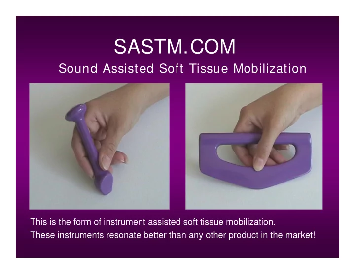
SLIDE 1 SASTM.COM
Sound Assisted Soft Tissue Mobilization
This is the form of instrument assisted soft tissue mobilization. These instruments resonate better than any other product in the market!
SLIDE 2
Our Philosophy:
It is our belief that working together each of the healing professions has a role to play in the patient achieving optimal health. Each excels in a unique way.
S.A.S.T.M. WILL ENHANCE YOUR SPECIALTY.
THE RESEARCH PROVES IT
SLIDE 3
CARPAL THERAPY INC. WOULD LIKE TO EXPRESS OUR GRATITUDE TO BALL STATE UNIVERSITY, THE BALL FAMILY, DOCTORS OF CENTRAL INDIANA SPORTS MEDICINE, THERAPISTS AND STAFF OF BALL MEMORIAL HOSPITAL, AND ALL WHO WERE AND ARE INVOLVED IN THE RESEARCH THAT HAS VALIDATED THE MODALITY OF I.A.S.T.M., AND HELPED EASE THE PAIN OF SO MANY.
SLIDE 4 Personal Clientele:
- Toyota
- BMW
- Ford
- Isuzu
- NFL (National Football League)
- NBA (National Basketball Association)
- MLB (Major League Baseball)
- USA Track & Field
- 2005 Summer Olympics
- For individual professional names visit sastm.com
- University athletic training programs
SLIDE 5
SECTION ONE
HI STORY AND RESEARCH OF I NSTRUMENT ASSI STED SOFT TI SSUE MOBI LI ZATI ON
SLIDE 6 1988 - History of I.A.S.T.M
- David sustained a total dislocation of the right knee while his was
training for a skiing competition. Surgery was needed to repair the medial and lateral ligaments. David received an ACL replacement, was left without a PCL, and the leg was casted. When the cast was removed, David had 0 range of motion in his knee.
- David learned through therapy how to perform soft tissue
mobilization and deep friction massage using his hands. After 6 months of treating himself, David was diagnosed with Bi-Lateral Carpal Tunnel and Trigger Finger resulting in surgery to his right hand.
SLIDE 7 1989 - Early Development of I.A.S.T.M. Instrument Assisted Soft Tissue Mobilization
Roller Concepts 1st Wooden 2nd Aluminum
Different size rollers were used for various pressures and various soft tissue being treated. David used the roller concept initially when treating his right hand after surgery. He continued to treat his left hand to prevent surgery.
SLIDE 8 1990 - One on One Fitness
- David specialized in training injured athletes as a Personal
Trainer using the Roller Concept and then developed the Instruments that fit body parts with handles and Weak-Link Training Concept.
- Dr. Tom Seiver saw the results first hand from one of his
- patients. The patient had a left frozen ankle. After 6
weeks of treatment with the instruments, an 80% range of motion was achieved.
- Case Study – Kevin Pugh, Canadian Ballet Dancer Study
was performed by David Graston, Andre Hall and Central Indiana Sports Medicine under the direction of Dr. Tom Seiver.
SLIDE 9 1990 - Early Development of I.A.S.T.M. Instrument Assisted Soft Tissue Mobilization
- The roller concept evolved into curvilinear instruments
with beveled edges acting as a roller for maximum
- coverage. The instruments also possessed a bladed edge
for separation and splaying of soft tissue. The instruments were designed using aluminum as the material.
Note: After David’s first treatment on his knee using these instruments, he gained a 30 degree range of motion.
SLIDE 10 1991 - Early Development of I.A.S.T.M. Instrument Assisted Soft Tissue Mobilization
- Aluminum material was replaced with stainless steel.
SLIDE 11 3 Patents – Original Technique
curvilinear instruments designed to fit the body parts being treated. Instruments possess 2 treatment edges (beveled & bladed).
- Beveled Edge was developed
from the roller concept and was designed to locate and break up adhesions.
- Bladed Edge was developed
to separate and splay the soft tissue after being located and broken up by the beveled edge.
SLIDE 12 1991 - Central Indiana Sports Medicine
- The success of a case study resulted in David
being recruited by Central Indiana Sport Medicine to open a clinic in Muncie, Indiana.
- Under the Direction of Dr. Tom Seiver, David
and collected outcomes to validate the Original Technique and Weak-Link Training.
SLIDE 13 1993 - Ball Memorial Hospital
- David went on to work with Ball Memorial
Hospital where they entered into a licensing and consulting agreement.
- They worked for a year doing IASTM research,
developing training and therapy protocols re: the Original Technique using the stainless steel instruments.
SLIDE 14 1994 - Ball Memorial Hospital Research
- Ball Memorial’s research on the
- riginal technique and weak link
research on athletes was collected by Ball State University’s Human Performance Lab.
SLIDE 15 1994 - Ball State University Human Performance Lab - IASTM Research
- Davidson CJ, Ganion L, Gehlsen G, Roepke J, Verhoestra B,
Sevier TL: Morphologic and functional changes in rat Achilles tendon following collagenase injury and GASTM. Journal of the American College of Sports Medicine 27 (5) 1995
- Sevier TL, Gehlsen GM, Wilson JK, Stover SA, and Helfst RH:
Traditional physical therapy vs. Graston augmented soft tissue mobilization in treatment of lateral epicondylitis. Journal of the American College of Sports Medicine 27 (5), 1995
SLIDE 16
Ball State University Human Performance Lab - IASTM Research
SLIDE 17
Ball State University Human Performance Lab - IASTM Research
SLIDE 18
Ball State University Human Performance Lab - IASTM Research
SLIDE 19 1994 - Physical Therapy & Training Center Opens
- Free standing Physical Therapy Clinic
utilizing the original Technique and tools in therapy.
- The original Technique and tools were
used with the Weak-Link Concept to increase performance in athletes of all ages by understanding that bio- mechanical imbalances were scar related.
SLIDE 20 1995 - Ball Memorial Hospital Opens Performance Dynamics
- The success of the clinic David opened in
Indianapolis, and differences in how to teach and market the product resulted in a mutual termination of relationship with Ball Memorial Hospital.
- Ball Memorial Hospital then moved their in
patient physical therapy to an outpatient setting known today as Performance Dynamics.
- Performance Dynamics developed and patented
their own curvilinear instruments under the direction of Dr. Tom Sevier.
SLIDE 21
1995 - Performance Dynamics
SLIDE 22 1997 - DAVID LEAVES CLINIC
- As a result of differences between the 3 original
partners regarding education, marketing and product development, the relationship was
- dissolved. David honored a 3 year non compete.
During this period, extensive research and development took place to develop a new method and instruments, and better teaching and treatment protocols.
SLIDE 23
1998 – GUA SHA
SLIDE 24 1998 – GUA SHA
scrape & SHA means ‘Sha-syndrome, or ‘reddish, elevated, millet-like skin rashes’ The technique of Gua Sha intentionally brings the Sha rash to the surface.
SLIDE 25
1998 – GUA SHA
SLIDE 26 1999 - Ball State University Human Performance Lab - IASTM Research
- Gehlsen GM, Ganion LR, Helfst RH. Fibroblast responses to
variation in soft tissue mobilization pressure. Medicine and Science in Sports and Exercise 31(4): 531-535, 1999
SLIDE 27 2000 - Carpal Therapy - SASTM.com
- 3 Years after David left (to
fulfill a noncompete agreement), Carpal Therapy, Inc. was opened in Sept. of 2000
research and development, manufactures and quality controls all products and seminars via SASTM.com.
SLIDE 28 Design
has been designed with a unique treatment edge based on a square surface concept.
levers thus increasing the mechanical advantage and reducing the effort and pressure needed from the clinician.
- Ceramic Polymer
- Straight Flat Treatment
Edge
SLIDE 29 How Does S.A.S.T.M. Work?
- Instruments aid in the location of restrictions via sound
waves and conform to the size of adhesion.
- Ergonomic design = Easily handled without excessive
gripping.
- Square Surface Design = Minimal pressure needed from
clinician to initiate controlled microtrauma in affected tissues without damaging healthy soft tissue.
- Microtrauma initiates reabsorption of excess scar tissue.
SLIDE 30 Inflammation
Inflammation is a necessary and important part of the treatment process. It is a natural response of the body that enables it to reabsorb and remodel scar tissue. However, uncontrolled inflammation allows fibrosis to thicken, restrict motion and create pain. We don’t treat areas that are painful, because we don’t want to inflame already inflamed tissue. We do want to create controlled inflammation. For that reason ice is an important last step. Ice decreases patient discomfort and recovery time.
SLIDE 31 Do symptoms accurately indicate the location of the cause of the problem?
i.e.: Should we treat the lateral epicondyle for Tennis Elbow? Inflammation creates a buildup of fibrosis.
Symptoms are not an accurate reflection of the location of the cause. They are the effect of a long standing, chronic problem. FOR A YOUNG ATHLETE, THE PAIN AT THE ELBOW IS THE EARLY SYMPTOMS WITH PROBABLE ADHESIONS LOCALIZED IN THE FOREARM. FOR THE MORE EXPERIENCE ATHLETE, THE INFLAMATIONAT THE ELBOW IS MORE CHRONIC, AND THEREFORE THERE IS INFLAMATION AND FIBROIS AT THE ELBOW, AND, A DIFFUSE ACCUMULATION OF FIBROSIS THROUGHOUT THE FOREARM. HE WILL ALSO HAVE AN INFILTRATION OF FIBROSIS IN THE DEEP TENDONS OF THE THUMB AND FOREFINGER FLEXORS AND EXTENSORS. Symptoms seldom occur at the start of a problem, unless trauma related. The deep thumb and forefinger extensors and flexors are usually the cause of lateral and medial epicondylitis!
SLIDE 32 SASTM.COM
Sound Assisted Soft Tissue Mobilization
This is the form of instrument assisted soft tissue mobilization. These instruments resonate better than any other product in the market!
SLIDE 33
SECTION THREE:
HAND HOLDS OF INSTRUMENTS AND EVALUATION AND TREATMENT STROKES
SLIDE 34 S.A.S.T.M. Hand Holds
Pencil Grip
Single Hand Hold Double Hand Hold
SLIDE 35 S.A.S.T.M. – Instrument Strokes
- Sweeping = Scanning (Lighter Pressure than
Treatment Pressure)
- Strumming = Treatment
- J Stroke = Treatment
Note: Any increase in treatment pressure should correspond with a decrease in treatment rate. To increase treatment pressure use a smaller instruments which has a smaller square surface rather than using more pressure.
SLIDE 36
Evaluation vs. Treatment
While using the S.A.S.T.M. instruments for soft tissue mobilization, there is a constant interchange between evaluation pressure and treatment pressure. Note: Evaluation or scanning pressure is less than treatment pressure.
SLIDE 37 FIND AN ADHESION AND DETERMINE BARRIER DIRECTION
- 1. LOCATE AN ADHESION BY SCANNING IN 4 DIRECTIONS.
2. WHICH OF THE 4 DIRECTIONS CREATES THE MAXIMUM VIBRATION / SOUND WAVE. THIS = THE BARRIER, WHICH IS THE HIGH SIDE OF THE ADHESION. THE BARRIER DIRECTION MAY CHANGE AS YOU TREAT DEEPER LAYERS.
RESCAN YOUR PALM FIND AN ADHESION DETERMINE THE DIRECTION OF ITS BARRIER
SLIDE 38 WHY DO WE TREAT?
SCARS ARE LIKE ONIONS. ONCE WE LOCATE THEM THROUGH SCANNING, WE CAN BREAK THEM DOWN, ONE LAYER AT A TIME. SOME OF THE TREATED TISSUE IS REABSORBED AND EXCRETED THROUGH THE KIDNEYS, AND SOME IS REMODELED. THE RESULT: WE HAVE TISSUE WITH LESS FIBROSIS, THAT IS MORE FUNCTIONAL. WE CONTINUE TO TREAT / REMODEL SCAR LAYERS UNTIL:
- A. THE PAIN IS ELIMINATED OR CONTROLED.
- B. FUNCTION IS IMPROVED
USUALLY 4-6 TREATMENTS YIELDS SIGNIFICANT IMPROVEMENT
SLIDE 39 4 WAYS TO INCREASE PRESSURE:
1 . DOW NSI ZE THE BLADE SI ZE
- EACH TI ME YOU DOW NSI ZE THE BLADE, SLOW DOW N
2 . CHANGE THE BLADE ANGLE:
- FROM 4 5 DEGREES TO 9 0 DEGREES
3 . I NCREASE THE STRETCH / CONTRACTI ON 4
ONLY PRESS HARDER AFTER YOU TRY THE FIRST 3. TRY EACH OF THESE
SLIDE 40 CROSS FIBER STROKES
AFTER TREATI NG THE HI GHEST BARRI ER, DO SEVERAL CROSS FI BER STROKES TO SEPARATE THE FI BERS OF THE I NVOLVED MUSCLE, TENDON OR LI GAMENT. 1 .) BARRI ERS OF SCARS I N THE TENDONS ARE USUALLY I N THE DI RECTI ON OF THE FI BERS OF THE TENDONS. 2 .) BARRI ERS OF SCARS I N THE MUSCLES CAN BE I N ANY DI RECTI ON. NOTE: I F THE DI RECTI ON OF YOUR BARRI ER STROKES AND CROSS FI BER STROKES ARE THE SAME, YOU CAN SKI P THE CROSS FI BER STROKES,BECAUSE YOU ACCOMPLI SH BOTH AT THE SAME TI ME.
SLIDE 41
TREATING LAYER DEPTH
SUPERFICIAL LAYERS: STRETCH OR CONTRACTED THE TISSUE. INTERMEDIATE LAYERS: SLIGHTLY RELAX THE TISSUE TO TREAT THIS NEXT LAYER. DEEP LAYERS: COMPLETELY RELAX THE TISSUE TO ALLOW THE INSTRUMENT TO SINK DEEP.
SLIDE 42 SASTM BASIC CONCEPT
- 1. SCAN 4 DIRECTIONS
- 2. TREAT IN DERECTION OF BARRIER
DIRECTION
- 3. FINISH CROSS FIBER IF NEEDED
- 4. WORK PROGRESSIVELY DEEPER
- 5. COMPLETE MOBILIZATION
SLIDE 43 BASIC USE OF S.A.S.T.M.
1.) WARM UP TISSUE TO TREAT:
- MOIST HEAT, STRETCH, U/ S, CARDIO
2.) TREAT 3.) MOBE / ADJUST 4.) EXERCISE / STRETCH 5.) YOU CAN INCLUDE ANY OTHER MODALITIES AT THIS POINT – THESE WON’T BREAK UP FIBROSIS HOWEVER.
- LASER ETC
- 6. ICE TO AREAS OF TREATMENT.
SLIDE 44
Patella Tendonitis Step 1: Patellar Ligament & Quad Tendon Superficial Layer
SLIDE 45
Patella Tendonitis Step 2: Ilio-Tibial Tract / I.T. Band Superficial Layer
SLIDE 46
Patella Tendonitis Step 3: Sartorius Tendon Superficial Layer
SLIDE 47
Patella Tendonitis Step 4: Gracilis & Medial Hamstrings Superficial To Intermediate Layer
SLIDE 48
Patella Tendonitis Step 5: Lateral Hamstring Superficial Layer