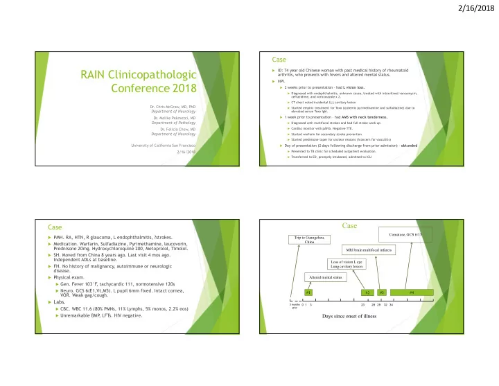
SLIDE 15 2/16/2018 15
Treatment of Balamuthia GAE
Only ~10 of 200 reported cases have survived
In some cases, survivors had complete return to normal
function without reported sequelae
Early diagnosis and treatment may increase odds of survival
CDC recommends combination therapy with:
- 1. Pentamidine 4mg/kg qD
- 2. Sulfadiazine 1.5g q6h (adults), 200mg/kg/day (peds)
- 3. Flucytosine 37.5 mg/kg q6h
- 4. Fluconazole 12 mg/kg/day
- 5. Azithromycin 20 mg/kg/day
- 6. Miltefosine 150mg daily (in US, only available through IND
filed by CDC) Duration of treatment. Several weeks to several
months/years.
Conclusions
Balamuthia mandrillaris is a free-living ameba
associated with granulomatous amebic encephalitis (GAE)
Affects immunocompetent patients more cases than rabies in the last 10 years! Diagnosis requires specialized testing Should contact CDC if suspicion is high (multifocal
encephalitis with negative studies)
Usually fatal, but 5% of patients have survived Treatment requires aggressive combination therapy
Thank you to all involved in this very challenging case
Dr. Barbara Haller – SFGH Microbiology Dr. Andrew Bollen – UCSF Neuropath Dr. Matt Wood – UCSF Neuropath Dr. Mike Reid – UCSF Infectious Disease Dr. Niraj Shanbhag – UCSF Neurology Dr. Mike Wilson – Derisi Lab Dr. Carole Glaser – DPH Many many others
For more information related to metagenomic deep sequencing, please visit: http://nextgendiagnostics.ucsf.edu
References
Baig AM. Pathogenesis of amoebic encephalitis:
Are the amoebae being credited to an 'inside job' done by the host immune response? Acta Trop. 2015;148:72-6. PMID: 25930186
CDC. Investigational drug available directly from CDC for the treatment of
infections with free-living amebae. MMWR (2013) 62;33:666
Detering H, et al. First Draft Genome Sequence of Balamuthia mandrillaris,
the Causative Agent of Amoebic Encephalitis. Genome Announc. 2015 24;3(5). PMID: 26404594
GreningerAL, et al. Clinical metagenomic identification of Balamuthia
mandrillaris encephalitis and assembly of the draft genome: the continuing case for reference genome sequencing. Genome Med. 2015;7:113. PMID: 26620704
Guarner J, et al. Histopathologic spectrum and immunohistochemical
diagnosis of amebic meningoencephalitis. Mod Pathol. 2007;20(12):1230-7. PMID: 17932496
Itoh K, et al. An autopsy case of Balamuthia mandrillaris amoebic
encephalitis, a rare emerging infectious disease, with a brief review of the cases reported in Japan. Neuropathology. 2015;35(1):64-9. PMID: 25186798
Khurana S, et al. Emergence of Balamuthia mandrillaris
meningoencephalitis in India. Indian J Med Microbiol. 2015;33(2):298-300. PMID: 25865989
Kiderlen AF
, et al. Assessment of Balamuthia mandrillaris-specific serum antibody concentrations by flow cytometry. Parasitol Res. 2009;104(3):663-
Kodet R, et al. Amebic encephalitis caused by Balamuthia mandrillaris in a
Czech child: description of the first case from Europe. Pathol Res Pract. 1998;194(6):423-9. PMID: 9689651
Latifi AR, et al. Presence of Balamuthia mandrillaris in hot springs from
Mazandaran province, northern Iran. Epidemiol Infect. 2016;144(11):2456-
Martínez AJ. Granulomatous amebic encephalitis: a review and report of a
spontaneous case from Venezuela. Acta Neuropathol. 1994;87(4):430-4. PMID: 8017178.
- Onyango CO, et al. Evaluation of a TaqMan Array Card for Detection of Central
Nervous System Infections. J Clin Microbiol. 2017;55(7):2035-2044. PMID: 28404679
- Qvarnstrom Y, et al. Multiplex real-time PCR assay for simultaneous detection of
Acanthamoeba spp., Balamuthia mandrillaris, and Naegleria fowleri. J Clin
- Microbiol. 2006;44(10):3589-95. PMID: 17021087
- Schuster FL. Cultivation of pathogenic and opportunistic free-living amebas. Clin
Microbiol Rev. 2002;15(3):342-54. Review. PMID: 12097243
- Schuster FL, et al. Balamuthia amebic encephalitis risk, Hispanic Americans.
Emerg Infect Dis. 2004;10(8):1510-2. PMID: 15503402
- Schuster FL, et al. Under the radar: balamuthia amebic encephalitis. Clin Infect
- Dis. 2009;48(7):879-87. PMID: 19236272
- Schuster FL, et al. Balamuthia mandrillaris, agent of amebic encephalitis:
detection of serum antibodies and antigenic similarity of isolates by enzyme
- immunoassay. J Eukaryot Microbiol. 2008;55(4):313-20. PMID: 18681845
- Tavares M, et al. Diagnosis of first case of Balamuthia amoebic encephalitis in
Portugal by immunofluorescence and PCR. J Clin Microbiol. 2006;44(7):2660-3. PMID: 16825409
- van der Beek NA, et al. Fatal Balamuthia mandrillaris Meningoencephalitis in the
Netherlands after Travel to The Gambia. Emerg Infect Dis. 2015;21(5):896-8. PMID: 25897644
- Visvesvara GS, et al. Leptomyxid ameba, a new agent of amebic
meningoencephalitis in humans and animals. J Clin Microbiol. 1990 28(12):2750-
- 6. Review. PMID: 2280005
- Wilson MR, et al. Diagnosing Balamuthia mandrillaris Encephalitis With
Metagenomic Deep Sequencing. Ann Neurol. 2015;78(5):722-30. PMID: 26290222.
- Yagi S, et al. Detection of Balamuthia mitochondrial 16S rRNA gene DNA in
clinical specimens by PCR. J Clin Microbiol. 2005;43(7):3192-7. PMID: 16000434