
SLIDE 1
Preventing Amputations In the Lower Extremities Of Patients With - - PowerPoint PPT Presentation
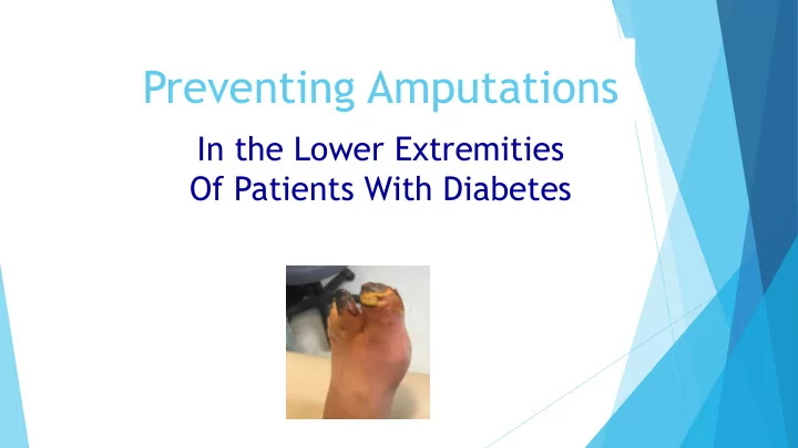
Preventing Amputations In the Lower Extremities Of Patients With Diabetes Several steps take place prior to the loss of a limb. The six steps are:- Diabetes, Neuropathy Ulceration Vascular disease Infection and
Several “steps” take place prior to the loss of a limb.
The six steps are:-
Diabetes, Neuropathy Ulceration Vascular disease Infection and amputation.
Each of these steps is preventable and one can take action to prevent the patient from escalating to the next step.
Treatment – Multifocal Approach
Thorough history Optimising glycaemic control Vascular supply ~ ABI = 0.45 referral Aggressive wound debridement Infection control Maintaining wound moisture control Appropriate offloading
Glycemic Control & vascular stasis
Control blood glucose -imperative healing chronic
wounds.
Hyperglycemia results – leukocyte dysfunction,
suppression lymphocytes.
Requires adequate tissue oxygenation = well vascularized wound bed = new granulation tissue
Smoking
Smoking greatest impact on PAD Cessation is the cornerstone of PAD treatment
Caution Debridement
Surgical debridement –
inappropriate for ulcers with vascular insufficient vascular supply –extreme Caution On patients on anticoagulants.
Emphasizing The Value Of Risk Stratification and Preventative measures.
Frequency visits depends on the
severity of the abnormality and the degree of intervention necessary to control ulcer risk.
Some hemorrhagic keratosis require
weekly, biweekly – monthly.
Debridement is extremely effective
preventing ulceration.
infection, hospitalization and
amputation.
Compromised sensory perception
L.O.P
.S – localized pressure, leading to tissue ischaemia and ulceration.
PN- high risk impaired balance and
gait.
Loss somatosensory afferents from
peripheral neuropathy =increased risk ulceration balance and gait control.
Initial Care for referred patient
Vascular - if pedal pulses are not
palpable , we order non – invasive arterial studies and obtain vascular consult based results.
Neurological exam. X-ray rule out osteomyelitis and assess
deformity that might be contributing to the wound.
Infection antibiotic management.
The Effects Of ESRD On Patients With Diabetes
Dialysis is an independent risk factor for
ulceration.
A 2x increase in the prevalence of other
lower extremity complications such as peripheral arterial disease (PAD) and amputations in dialysis-treated patients.
Found an increase in foot ulcerations in
patients with ESRD.
A 4X increase in diabetic foot
complications, defined as infection, gangrene and amputation.
End-stage renal disease (ESRD)
Kidney disease increases the risk of
peripheral arterial disease (PAD) 3X in comparison to patients without renal disease but the severity of PAD worsens as kidney disease progresses.
Calciphylaxis is a thrombolytic event
that provokes ischaemia and tissue infarction.
Common lower extremities. Begin painful red areas that develop
into indurated plaques followed by eschar, ulceration and gangrene.
One year mortality rate > 50% often
2nd to sepsis deriving ulcers.
Clinical (for osteomyelitis) ➢ History: long wound duration, recurrent infection ➢ Exam deep large(>2cm2) ulcer, bony prominence, visible bone/joint, “sausage” toe ➢ Probe-to bone: useful if done and interpreted correctly ➢ Blood tests: WBC, ESR, C-RP , ? Biomarkers
Clinical Classification Diabetic Foot Infection
Clinical Manifestations* IDSA Severity
IWGDF PEDIS
No purulence or inflammation (erythema, pain, warmth, tenderness, or induration) Uninfected 1 Infected(>=signs/sx inflammation) But erythema ,=2cm around ulcer, infection limited to skin or superficial subcutaneous tissues Mild 2 >=1 of following: cellulitis>2cm Lymphangitis; subQ spread Deep abscess; gangrene; Muscle, tendon, joint or bone involved Moderate 3 Systematic toxicity or metabolic instability Severe 4
➢ Atrophy of the short extensor muscle. ➢ Atrophy of the intrinsic muscles of the arch. ➢ Hammer toe deformities ➢ Hallux valgus deformity ➢ Gait instability ➢ Falls
Diabetic Motor Neuropathy
Charcot feet – heel walk – cannot raise toes Tibilas anterior weakness- Foot slap
Inactive Charcot Foot
When there is no inflamation it is inactive
Thermography
Diagnosis of Charcot's foot
is supported, where available, by the use of thermography, which will show a skin temperature
contralateral foot.
Early Active Charcot
Misdiagnosis Cellulitis, Gout, Deep venous
thrombosis.
Evaluating Equinus
Silfverskiold Test
Equinus
Equinus – the most profound casual agent in foot
pathomechanics
Life threatening significantly increases risk of diabetic foot ulcer
Refer orthopaedic surgeon Diabetic foot clinic
Equinus Treatment
Debridement wound Offloading – moonboot Tendo-achilles lengthening to heal a diabetic fore-foot ulcer Refer orthopaedic surgeon for surgery options Conservative prior ulceration – manual stretching – night
splints
Equinus Treatment
Neuropathic Diabetic Wound
One should initially consider the
“VIPs” (vascular, infection and pressure).
Increased plantar pressure is a
common reason for non-healing of
Diabetic neuropathic wound
Damaged nerve impulses
control muscles ie motor nerves.
Pain , touch or positional
sense ie sensory nerves.
As a result of peripheral
neuropathy they may develop
increased risk of falling.
An ankle foot (AFO) or orthotics with
extra – depth shoe can be appropriate in some cases
Meticulous wound management,
including debridement. Vascular surgeon consult – revascularization.
The knee walker scooter moonboot. AFO – orthotics modification remains
healed.
This is due to loss of plantarflexory function of the gastrocnemius muscle and subsequent overload at the plantar heel in gait.
Ulceration - treatment
Digital amputation significant indicator
Loss digits alternation of
resulting in changes pressure location new areas osseous prominence >PRESSURE – ulceration –infection AMPUTATION.
Multiple hospitalizations and
re –operations
Preventing Diabetic foot Recurrence
After achieving healing
Appropriate shoe gear Orthotics or bracing to help prevent recurrence Therapeutic footwear in those with severe foot deformity Refer surgeon Distal toes tenotomy Charcot reconstruction Achilles lengthening
I frequently get orthotics to get rocker soled shoes, metatarsal pads and accommodation under the affected areas.
Emphasizing appropriate Shoegear And Patient Education
Evaluation and management of
minor trauma triggers like foot deformity, pressure callus and undetected injury may prevent amputation
Encourage compliance with
diabetes control
Emphasize the importance of
visual foot exams at home.
Emphasizing appropriate Shoegear And Patient Education
Pressure relieving shoes and
amputation
Educate patients every visit Explain the potential impact
Current interventions to address gait and balance diabetic peripheral neuropathy
Physiotherapy – guided training Postural control training Custom insoles – enhance balance
control in individuals with neuropathy. There is a need to improve, restore or replace inputs regarding plantar pressure proprioception to
improve the motor control of gait and balance for patients to walk safely.
SurroGait Rx
Wearable technology has
a potential benefit high – risk population.
Treatment
Offloading the wound. Surgical shoes Casts TCC Crutches Walkers Wheelchairs
Flexor tenotomy – distal tip toes diabetic neuropathy
A full thickness ulcer 4x6mm, a slight hyperkeratotic
rim with red granular base positive probing bone
Radiographic findings cortical disruption -concern
Oral antibiotics started. The triad of diabetic neuropathy Hammertoe deformity and repetitive trauma resulted
ulceration in this patient
Digital amputation most common foot amputation –
eradicate infection
Subungual squamous cell carcinoma of the nail bed.
Presentation fingernail and a linear
pigmented streak below right hallux nail plate.
Dermatologist review – following day
placed dermatology clinic.
Review radiographs for underlying
Subungual squamous cell carcinoma of the nail bed.
Nail plate avulsion and 3mm punch biopsy.
This case study remains under review as nail bed abnormality high risk non- healing with her diabetes and confirmation of pathology dermatologist – benign. In discussion dermatologist high risk – rerefer Urgent review change pigment change nail matrix.
Level pain, presence infection, erythema ,
edema, granulation tissue and drainage. Risk
–soft tissue infection and osteomyelitis of distal phalanx may occur.
Treatment – prophylactic antibiotic cover.
Pedal pulses, resolve infection prior treatment.
Conservative treatment failed review
patient history. Diabetes,pvd,meds,
Conclusion
Research has shown that multidisciplinary teamwork,
the addition of a podiatry service, prescription footwear and home temperature monitoring can prevent diabetic foot ulcers and amputation.
Prevention of foot complications in diabetes is key in
improving the patient’s quality of life, reducing mortality and lowering healthcare costs.
Thank-you - Jacqui Journeaux