
SLIDE 1
Rasmus Bro
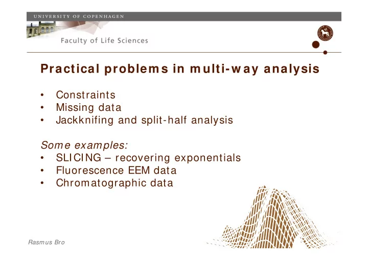
Practical problem s in m ulti-w ay analysis
- Constraints
- Missing data
- Jackknifing and split-half analysis
Some examples:
- SLICING – recovering exponentials
- Fluorescence EEM data
- Chromatographic data