
SLIDE 1
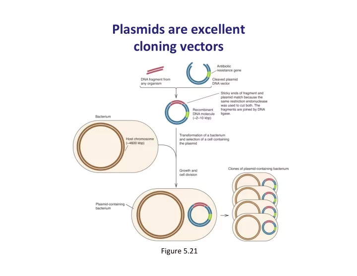
Plasmids ¡are ¡excellent ¡ ¡ cloning ¡vectors ¡
Figure ¡5.21 ¡
Plasmids are excellent cloning vectors Figure 5.21 - - PowerPoint PPT Presentation
Plasmids are excellent cloning vectors Figure 5.21 Figure 11.11 Order of restriction enzyme cut sites in polylinker Ampicillin lacZ resistance Apo I - Eco RI Ban II - Sac I Acc 651 - Kpn I Ava
Figure ¡5.21 ¡
Figure 11.11
Ampicillin resistance Polylinker Origin of DNA replication
2686 base pairs lacI lacZʹ″ ʹ″
ApoI - EcoRI BanII - SacI Acc651 - KpnI AvaI - BsoBI - SmaI - XmaI BamHI XbaI AccI - HincII - SalI BspMI - BfuAI SbfI PstI SphI HindIII
Figure 11.12
AmpR lacZʹ″ ʹ″ Vector Foreign DNA Opened vector Recyclized vector without insert Vector plus foreign DNA insert Transformants blue (β-galactosidase active) Transformants white (β-galactosidase inactive)
Digestion with restriction enzyme Join with DNA ligase Transform into Escherichia coli and select on ampicillin plates containing Xgal
Figure 11.18
Capsid genes cos att int xis J N QSR cos Replaceable region
(replacement vector)
(insertional vector) β-Gal gene β-Gal gene Another substitution Another substitution
Figure 11.19 Replaceable region Infective lambda virion R R R R L L L L cos
Foreign DNA Hybrid DNA
Digestion with restriction enzymes Ligation with foreign DNA Packaging cloned DNA into phage head Phage assembly
Figure 11.13
Well-developed genetics Many strains available Best known bacterium Easily transformed Nonpathogenic Naturally secretes proteins Endospore formation simplifies culture Well-developed genetics Nonpathogenic Can process mRNA and proteins Easy to grow Potentially pathogenic Periplasm traps proteins Genetically unstable Genetics less developed than in E. coli Plasmids unstable Will not replicate most bacterial plasmids Advantages Disadvantages Escherichia coli Bacillus subtilis Saccharomyces cerevisiae
Figure 11.15
Ampicillin resistance Promoter ESM CEN Promoter Polylinker (cloning site)
t/pa
t/pa
Figure 11.20
Polylinker M13 genomic DNA lacP lacZʹ″ ʹ″ Phage in clear plaques have cloned DNA Phage in blue plaques do not have cloned DNA
EcoRI KpnI XbaI SalI PstI HindIII SmaI BamHI
Figure ¡5.18ab ¡
Photo ¡courtesy ¡of ¡FOTODYNE ¡Incorporated. ¡
Figure ¡5.19ab ¡
Figure 11.5
Foreign DNA Sticky ends Vector Cloned DNA Introduction of recombinant vector into a host Cut with restriction enzyme Add vector cut with same restriction enzyme Add DNA ligase to form recombinant molecules
Figure 11.9
Figure ¡5.45 ¡
Figure 11.14
Plunger Helium gas Gas vent Disc Microprojectiles with transfecting nucleic acid Fine screen Rough screen Target tissue
Figure 11.6 Transformant colonies growing on agar surface X-ray film Positive colonies Replica-plate onto membrane filter Partially lyse cells; add specific antibody; add agent to detect bound antibody in radiolabeled form Lyse bacteria and denature DNA; add RNA or DNA probe (radioactive); wash
Autoradiograph to detect radioactivity
Figure ¡5.31 ¡
Figure ¡5.22 ¡