outline
play

Outline Office Procedures in Orthopaedics Knee exam Knee - PowerPoint PPT Presentation

8/7/2013 Physical Exam Skills and Outline Office Procedures in Orthopaedics Knee exam Knee aspiration and injection Shoulder exam Subacromial bursa injection UCSF Essentials of Primary Care August 14, 2012 Carlin Senter, M.D.


  1. 8/7/2013 Physical Exam Skills and Outline Office Procedures in Orthopaedics • Knee exam • Knee aspiration and injection • Shoulder exam • Subacromial bursa injection UCSF Essentials of Primary Care August 14, 2012 Carlin Senter, M.D. 
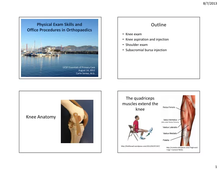
The quadriceps muscles extend the knee Knee Anatomy http://thefitcoach.wordpress.com/2012/04/07/267/ http://scientia.wikispaces.com/Thigh+and +Leg+-+Lecture+Notes 1

  2. 8/7/2013 The quadriceps muscles merge to form the The hamstrings flex the knee quadriceps tendon… patellar tendon www.hep2go.com There are 4 Pes anserine bursa main ligaments in the knee http://meded.ucsd.edu/clinicalmed/joints.htm 2

  3. 8/7/2013 Meniscus Knee exam Common Causes of Knee Pain by Location of Musculoskeletal work-up Symptoms • H istory • Anterior: • Medial - Medial joint-line: meniscus - Patellofemoral syndrome tear or OA - Quadriceps tendinitis • I nspection - MCL sprain - Patellar tendinitis - Pes anserine bursitis • P alpation • Lateral: • Posterior - Lateral jointline: meniscus tear - Hamstring tendinitis or OA - Gastrocnemius strain • R ange of motion - IT band syndrome - OA, meniscus tears, - LCL sprain (rare) effusion, popliteal cyst…. - Fibular head: fracture (rare) • O ther T ests 3

  4. 8/7/2013 Palpation of joint line Inspection seated or supine http://www.rheumors.com/kneeexam/palpation.html http://doctorhoang.wordpress.com/20 http://meded.ucsd.edu/cl 10/09/06/valgus-knee-and-bunion/ inicalmed/joints.htm Palpation of patella - supine Palpation of patellar facet Ballottement 4

  5. 8/7/2013 Knee range of motion Other Tests: Lachman to evaluate ACL Sensitivity 75-100% Specificity 95-100% • ROM: normal 0-135 – Determine if knee is locking or if ROM is limited due to effusion – Locking: think bucket handle meniscus. • Urgent xrays, MRI • Urgent referral to sports surgeon for arthroscopy Permission for use provided by Magee, DJ. Orthopaedic Physical Assessment, 5 th ed. 2008. Dr. Charles Goldberg, UCSD MCL and LCL PCL: Posterior Drawer 5

  6. 8/7/2013 Meniscus: McMurray Meniscus: Thessaly Sensitivity medial 65%, Specificity medial 93% Magee, DJ. Orthopaedic Physical Assessment, 5 th ed. 2008. Meniscus: Squat Knee exam practice • Standing: inspection • Supine – Varus or valgus – Patellar facets • Sitting: palpation – Patellar grind – ROM – Joint line – Special tests – Femoral condyles • Lachman – Tibial plateau • Posterior drawer – Fibular head • Varus 0 and 30 • Valgus 0 and 30 • McMurray medial and lateral • Thessaly • Squat 6

  7. 8/7/2013 Intra-articular corticosteroid injections: do they work for knee OA? • Good short-term pain relief – Effect size 0.72 at 2 and 3 weeks • No significant effect on function – Effect size 0.06 Knee aspiration and injection • No evidence for long-term pain relief • Clinical effect independent of degree of inflammation present – Don’t need to restrict injection just to those with effusion • Frequency: general practice once every 3 months max – Concern for cartilage toxicity with more than 4/year • AAOS: recommends for short-term pain relief (level II) Zhang W et al. OARSI recommendations for the management of hip and knee osteoarthritis: Osteoarthritis Cartilage. 2010 Apr;18(4):476-99. Superolateral approach Injection set-up bucket • Patient supine • Betadine • Extend knee • Ethyl chloride • Bump under knee so • Alcohol swabs • 4x4 guaze flexed 10-20 degrees • Superior border patella • Bandaids • Lateral border patella • 1cm below • Mark with syringe cover or tip of pen 7

  8. 8/7/2013 Injection prep Needles, syringes, meds Why use local anesthetic with steroid Corticosteroids injection? • Dilute the steroid – Decrease likelihood of steroid atrophy – Decrease irritant nature of steroid crystals causing post-injection flare • Pain relief – Diagnostic and therapeutic (subacromial more than knee) • Floculation: combining steroid and local anesthetic can precipitate crystals. Carefully inspect for precipitate before injection. 8

  9. 8/7/2013 Why aspirate the effusion before Aspiration injection? • Clinically – Decreased pain and stiffness because effusion gone – More effect of steroid because not diluted by effusion – Inspect fluid for inflammation/infection, send to lab if question – Confirms that injxn was intra-articular • Significantly greater improvement in VAS for patients who had joint aspirated at time of injection in knee OA patients (Gaffney K et al, Ann Rheum Dis, 1995.) • Reduction in relapse for 6 months after injection in RA patients (Weitoft T et al, Ann Rheum Dis, 2000.) Post-injection patient instructions Contraindications to steroid injection • Rest: no definitive evidence-based • Joint infection • Fracture recommendation – Recommendations in literature vary • Prosthetic joint • No restrictions • Hemarthrosis (theoretically higher risk of • Bed rest x 24 hours infection) • Light activity x 7 days, no weight bearing exercise • Soft tissue infection overlying joint • Avoid swimming, hot tub, bath x 24 hours – Let injection site heal 9

  10. 8/7/2013 Relative contraindications to steroid Risks of steroid injection in the knee injection • • Corticosteroid injection within past 4 months Diabetics: increased blood sugar, 300 mg/dl starting as early as 2 hours after, lasting 5 days • Suppression of hypothalamic pituitary adrenal axis, mild • Coagulopathy (ok if on warfarin but check – Lasts 1-3 days post-injection • Facial flushing: 10% with Kenalog recent INR, make sure not >> 3) – 19-36 hours post-injection • • Poorly controlled diabetes Skin or fat atrophy • Post-injection steroid flare: 1-10% – Synovitis in response to injected crystals – Within hours - 48 hours post-injection – More common in soft tissue injections (20% of trigger points) than intra- articular injections • Septic arthritis: 1/3000-1/50,000 – 1-2 days after injection • Possible risk of chondrocyte toxicity with repeated injections Habib GS. Clin Rheumatol, 2009. UpToDate, “Joint aspiration or injection in adults,” 2010. My current knee injection steps Knee injection 1. Patient supine with bump under knee 2. Mark injection site (superior lateral) 3. Betadine x 3 4. Alcohol x 1 5. Ethyl chloride for skin anesthesia 6. Alcohol again 7. 22g needle attached to 10cc syringe containing 5cc of 1% lidocaine without epi 8. Slowly advance and inject lidocaine, 1mm at a time 9. Feel resistance give when in joint 10. Aspirate, make sure fluid straw-colored and clear 11. Keep needle in place, switch syringe 12. Inject 1cc of 40mg kenalog 10

  11. 8/7/2013 Underlying Anatomy - Bones Shoulder anatomy Acromion Greater Tuberosity • Humerus Clavicle • Scapula o Glenoid o Acromion o Coracoid o Scapular body • Clavicle • Sternum Glenohumeral Joint Lesser Tuberosity The LABRUM is a fibrocartilaginous ring of tissue that attaches to the glenoid rim & deepens the glenoid fossa Spine of Acromion scapula is at the level of T3 Bottom of scapula is at level of T7 11

  12. 8/7/2013 Supraspinatus (Abduction ) The Rotator Cuff Muscles (SITS) The tendons of the rotator cuff muscles reinforce the capsule Greater Posterior of the glenohumeral Tubersosity View joint. Lesser Tuberosity Infraspinatus (External rotation)) Anterior View Teres Minor Subscapularis (External rotation) (Internal Rotation) The Biceps Muscle Shoulder exam • #1 Supination of the elbow (screwing, twisting) • #2 Flexion of the elbow Long head 3 attachments: • Radial tuberosity (distal) • Glenoid (long head) • Coracoid (short head) Short head 12

  13. 8/7/2013 Cervical Spine Neck examination Spurling’s Maneuver • Neck extended • Inspection • Head rotated toward • Palpate CS affected shoulder • FF and extension • Axial load placed on the cervical spine • Spurlings • Reproduction of patient’s shoulder/arm pain indicates possible nerve root compression Shoulder examination Shoulder examination • Inspection • Inspection • Palpation – Patient in gown • Palpation • ROM • ROM http://meded.ucsd.edu/clinic • Strength almed/joints2.htm, • Strength permission granted by Dr. – Supraspinatus Charles Goldberg, UCSD SOM – Supra – Infraspinatus & – Infra and teres Teres minor minor – Subscapularis – Subscapularis • Other tests • Other tests http://meded.ucsd.edu/clinicalmed/joints 2.htm, permission granted by Dr. Charles Goldberg, UCSD SOM 13

  14. 8/7/2013 Range of motion Range of motion Internal rotation Abduction External rotation Flexion Supine shoulder PROM Other tests • Rotator cuff strength • Impingement tests • Biceps • Labrum • AC joint 14

  15. 8/7/2013 Infraspinatus and teres minor = Supraspinatus = abduction external rotation Supraspinatus Infraspinatus Teres minor Empty can Photos from Dr. Christina Allen Photos from Dr. Christina Allen Subscapularis = internal rotation Subscapularis = internal rotation Subscapularis Subscapularis Lift-Off Belly press Photos from Dr. Christina Allen Photos from Dr. Christina Allen 15

Download Presentation
Download Policy: The content available on the website is offered to you 'AS IS' for your personal information and use only. It cannot be commercialized, licensed, or distributed on other websites without prior consent from the author. To download a presentation, simply click this link. If you encounter any difficulties during the download process, it's possible that the publisher has removed the file from their server.

Recommend


More recommend